New Hormone Treatment Rescues Cognition in Down Syndrome Patients
Almost everyone has come across a person affected by Down syndrome, which is no surprise considering it’s the most common cause of genetic intellectual disability. Because of its chromosomal roots, the disorder is difficult to target and impacts brain and body development dramatically. Fortunately, a recent breakthrough from the University of Lille may reveal an exciting new treatment option to restore some cognitive capabilities in Down syndrome patients through the non-invasive administration of a hormone called GnRH.
🧠Article: “GnRH replacement rescues cognition in Down syndrome” (09/2/2022) - Manfredi-Lozano, M., Leysen, V., Adamo, M., Paiva, I., Rovera, R., Pignat, J.M., Timzoura, F.E., Candlish, M., Eddarkaoui, S., Malone, S.A. and Silva, M.S., 2022. GnRH replacement rescues cognition in Down syndrome. Science, 377(6610), p.eabq4515.
🧠Introduction and Methods:
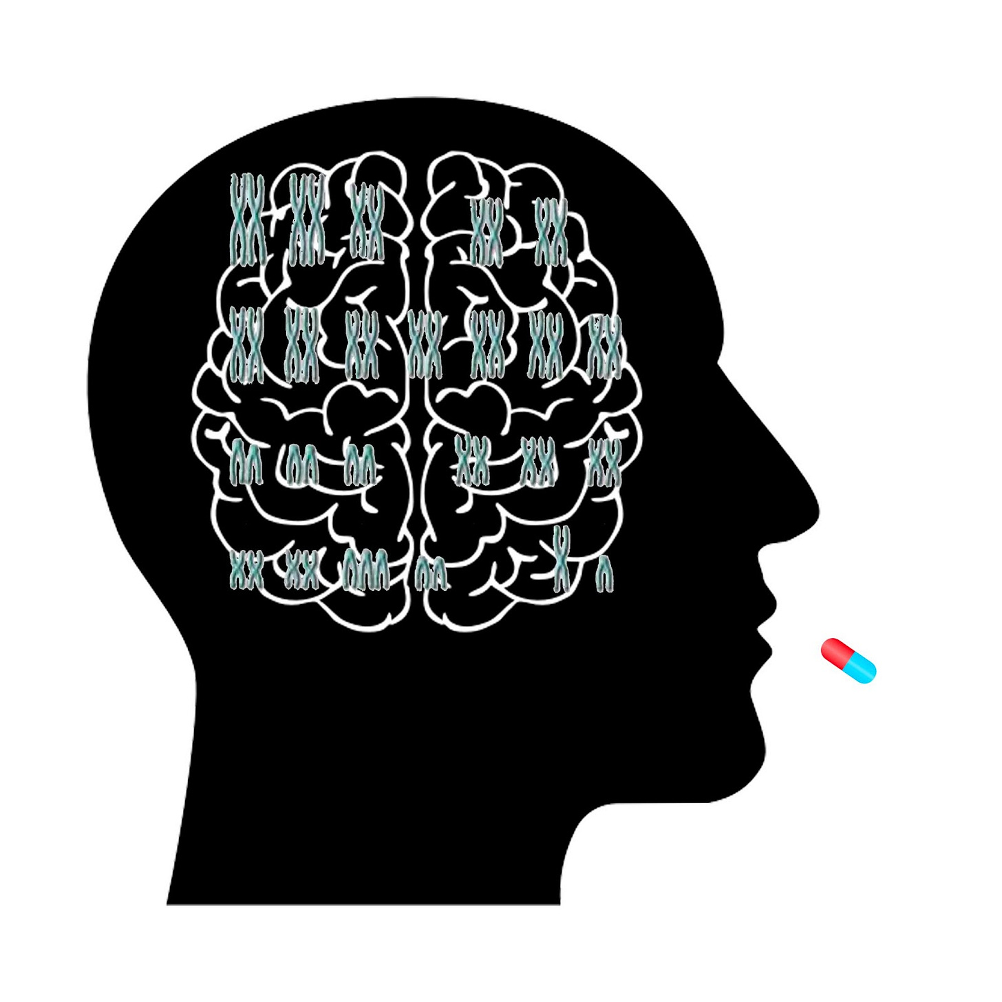
Down syndrome is a genetic disability that arises when an individual is born with three chromosomes in the 21st position instead of two. This additional chromosome results in abnormal gene expression which can cause a loss of smell, a change in physical appearance, infertility, and a neurodegenerative loss of cognition in a form that mirrors early-onset Alzheimer’s disease.
University of Lille researchers noticed that infertility and loss of smell are both symptoms of a deficiency GnRH (gonadotropin-releasing hormone), a hypothalamic hormone involved in activating sex steroids, and they wondered if this mechanism may be linked to Down syndrome. The team decided to administer a GnRH to human Down syndrome patients and mice that expressed a Down syndrome phenotype to see if cognition and olfaction could be improved. They used immunohistofluorescence to identify GnRH neurons in mouse brains and ran a series of behavioral tests to evaluate cognitive improvements in both mice and humans after a hormone treatment protocol.
🧠Results and Discussion:
In mouse models, a 3-D analysis of brain tissue showed that the Down syndrome-analogous cognitive impairments, which usually progress around the time of puberty, coincided with a loss of GnRH neurons in the hypothalamus. Although the hormone’s role was previously thought to be almost exclusively reproductive, the researchers found that GnRH neurons extended into areas that controlled cognition and social behavior, and they even found some in the cerebral cortex which controls intelligence and personality.
Furthermore, an injection of GnRH cells into the afflicted mice fully reversed the cognitive impairments and loss of smell. Though infertility was not improved, the massive gains in visuospatial memory and object recognition warranted a pilot study on human Down syndrome patients. Seven subjects were given the Montreal Cognitive Assessment to measure executive function, attention, and memory, all of which were at a deficit. An fMRI also revealed highly impaired connectivity between brain regions. The patients were then fixed with an external pump that released GnRH every two hours, mimicking the secretion process of the hypothalamus. The result was a significant improvement across all areas measured on the cognitive assessment and a dramatic enhancement of brain connectivity. The main connectivity improvements were seen in the visual, sensorimotor, and prefrontal cortices.
Ultimately, this study didn’t just reveal the important cognitive role of a hormone that was thought to be exclusive to reproduction, but it also opened the door to an exciting new treatment path for a disease that currently has no pharmacological interventions. This looks to be an exciting time for Down syndrome research, with the next few years hopefully offering some relief for those who suffer from cognitive impairments as a result of the disease.
A Newly Discovered Type of Synapse Controls Your DNA
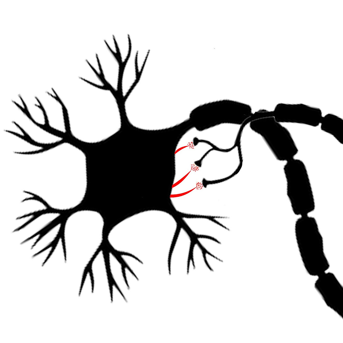
When it comes to communication between neurons, the synapse is where the magic happens. This tiny gap between the axon terminal of one nerve cell and the dendrite of another is where chemical neurotransmitters are released to influence whether or not the receiving cell will fire an action potential. Since the synapse is so elemental when it comes to brain function, researchers from the Howard Hughes Medical Center were excited to find that, hiding stealthily in the hair cells on mouse neurons, was a type of synapse that had never been identified before.
🧠Article: “A serotonergic axon-cilium synapse drives nuclear signaling to alter chromatin accessibility” (9/1/2022) - Shu-Hsien Sheu, Srigokul Upadhyayula, Vincent Dupuy, et. al. A serotonergic axon-cilium synapse drives nuclear signaling to alter chromatin accessibility, Cell,Volume 185, Issue 18, 2022, Pages 3390-3407.e18, ISSN 0092-8674, https://doi.org/10.1016/j.cell.2022.07.026.
🧠Introduction and Methods:
Outside of their role in embryonic development, the function of primary cilia has long been somewhat of a mystery. They have been shown to play a role in the Sonic hedgehog (Shh) system which affects developmental cell growth and differentiation, but these tiny hairs tend to disappear in most mature cell types. However, for reasons that were previously obscure, primary cilia remain attached to neurons and glial cells across the lifespan.
Research has demonstrated that these structures contain receptors that respond to neurotransmitters like dopamine and serotonin, and that removing them causes minor impairments to cognition, but their functional role in adult neurons remained mostly unknown. To shed light on this, the research team out of Howard Hughes used antibody staining and electron microscopy to examine the primary cilia of neurons in the hippocampus and raphe nuclei. They later engineered a cilia-targeted sensor to check for the release of serotonin around the hair cell.
🧠Results and Discussion:
High-powered microscopy revealed that the primary cilia extending from neuronal cell bodies actually serve as chemical synapses communicating with the axons of neighboring neurons. They found that the cilia contain vesicles (tiny transport sacs that carry neurotransmitters) as well as 5-HTR6 serotonin receptors and other synaptic structures that allow for intercellular communication. Nothing resembling this “axo-ciliary synapse” had ever been discovered previously.
Furthermore, by pulsing serotonin onto these ciliary receptors the researchers found that they could affect a pathway that produces RhoA, a protein involved in determining cell morphology. By altering this pathway the team showed that the axon-ciliary synapse modulated how tightly DNA was wound, determining whether certain genes could be translated into proteins and affecting the function of the hippocampus as a whole. Therefore, the study not only solved the mystery of the primary cilia and revealed a brand new kind of synapse, it also discovered an important cellular mechanism that could affect our understanding of certain cognitive disabilities.
What Science Says About Your Lizard Brain
If you’ve ever blamed an irrational decision on your “lizard brain,” you’re following the tradition of neuroscientist Paul MacLean. MacLean proposed the triune brain model in the 1970s, asserting (with little empirical backing) that the human brain had a survival-oriented reptilian core upon which the more complex limbic system and neocortex evolved. Modern neuroscientists have come to regard MacLean’s theory as an annoyingly enduring myth, though a recent publication from Germany’s Max Planck Institute for Brain Research shows that the comparison between mammalian and reptilian brains is worth examining.
🧠Article: “Molecular diversity and evolution of neuron types in the amniote brain” (9/2/22) - Hain D, Gallego-Flores T, Klinkmann M, Macias A, Ciirdaeva E, Arends A, Thum C, Tushev G, Kretschmer F, Tosches MA, Laurent G. Molecular diversity and evolution of neuron types in the amniote brain. Science. 2022 Sep 2;377(6610):eabp8202. doi: 10.1126/science.abp8202. Epub 2022 Sep 2. PMID: 36048944.
🧠Introduction and Methods:
Over 300 million years ago, the evolutionary paths of mammals and reptiles diverged from one another. Despite this ancient branching there are distinct neural similarities between the classes, including the general structural arrangement of the forebrain, midbrain, and hindbrain. It’s not obvious that this would be the case considering that other organisms, such as mollusks, developed a vastly different neural architecture in the same evolutionary time span. The disconnect between the distant evolutionary divergence and seemingly similar brain structure between reptiles and mammals piqued the interest of scientists out of Max Planck.
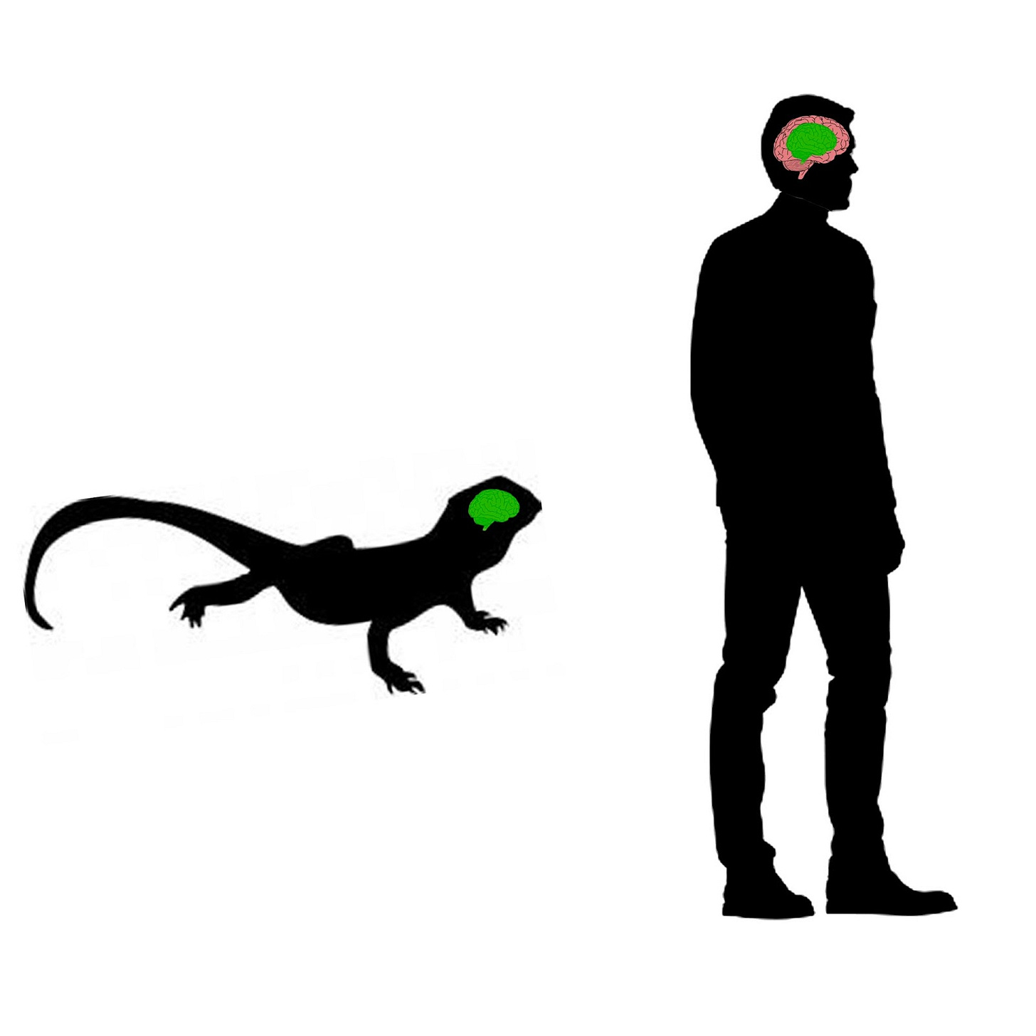
The researchers were interested in understanding whether the system of neural differentiation into specific subregions (basal ganglia, cerebral cortex, thalamus, etc.) were inherited from a shared ancestor hundreds of millions of years ago, or if reptiles and mammals converged independently onto a similar result. To uncover this, the team performed a genetic analysis on the brains of one reptile and one mammal, an Australian bearded dragon and a mouse (often chosen for its human-like neural structure). They performed single-cell RNA sequencing on 89,015 neurons throughout the bearded dragon brain and classified them under 11 different brain regions, and did the same for 70,968 mouse cells.
🧠Results and Discussion:
The Planck team ultimately uncovered a surprising degree of genetic conservation between reptile and mammal brains. Through transcriptional analysis, they found that the general neuron classes that mark distinct subregions of the brain were largely the same across species, indicating that this differentiation system is a remnant from an ancient common ancestor. In other words, it appears that there was a primitive creature roaming the earth over 300 million years ago with a similar brain architecture to that of modern mammals and reptiles. Though reptile and mammal neurons could be grouped into the same general categories, the form of the neurons themselves did diverge significantly in response to different evolutionary pressures.
Of course, there is still no evidence for MacLean’s claim that the mammal brain consists of additional layers on top of a reptilian brain. In fact, since the team found that even complex structures like the cerebral cortex were conserved, it refutes the idea that certain brain regions are more ancient than others. The reality is that both mammals and reptiles can thank a single primitive ancestor for passing on much of the brain organization we see today, which might be even more amazing than the triune model.


