

The Neural Newsletter for 8/23 - 8/30
The Neural Newsletter for 8/23 - 8/30
🧠 Last Week in Neuroscience


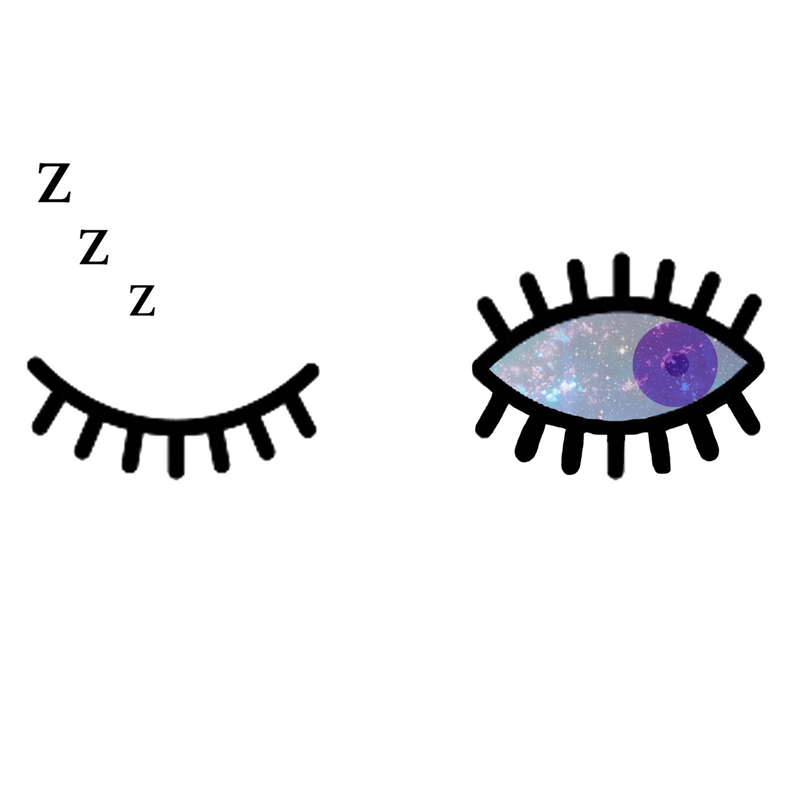
Eye Movements During REM Sleep are Surveying Your Dreamworld:
Few cognitive processes are as intriguing and mysterious as dreaming. Scientists have always been eager to find objective measurements that could help us better understand the bizarre and revealing performances our brains display at night, but most of the big questions remain unanswered. We do know that our most vivid dreams come during the period known as REM (rapid eye movement) sleep, which is also linked to learning and emotional regulation. The fitful eye movements that characterize this phase have been somewhat of a mystery, with many researchers writing them off as the product of random brainstem activity. This past week, however, researchers from the University of California, San Diego made a discovery about this activity which suggests that they are far from random, and may actually reveal a lot about our subjective experience during sleep.
🧠 Article: “A cognitive process occurring during sleep is revealed by rapid eye movements” (August 25, 2022) - Senzai, Y., & Scanziani, M. (2022). A cognitive process occurring during sleep is revealed by rapid eye movements. Science, 377(6609), 999–1004. https://doi.org/10.1126/science.abp8852
🧠 Introduction and Methods:
Though it’s known that REM sleep is linked to dreaming, there are conflicting hypotheses about the role and cause of rapid eye movements. Much of this uncertainty stems from the fact that previous studies have relied on human subjects reporting on their dreams with no objective measurement of cognitive processes. The UCSF team sought to eliminate any subjectivity by tracking concrete neural activity linked to rapid eye movement in rats during REM sleep.
To accomplish this, the team monitored head direction (HD) neurons in the mice’s anterodorsal nucleus of the hypothalamus during REM sleep. These cells track the orientation of the head as a waking animal traverses its environment. The researchers used extracellular probes to monitor HD cell activity while tracking REM phase eye movements using cameras (since mice sometimes open their eyes during sleep). They recorded the activity of HD cells in six mice, examining between 30 and 72 cells in each.
🧠 Results and Discussion:
Using their cameras, the team found that the eye movements during REM sleep were similar to the saccadic eye movements that animals use to survey their environment during waking. They also found that the HD neurons were firing in patterns that resembled the patterns that were expressed when mice were exploring new terrains. Even though the sleeping mouse’s head remained in a fixed position the brain perceived the head as moving in step with the eye movements of REM sleep, demonstrating a coupled pattern that mirrored brain activity in waking and active mice. They even used an algorithm to show that leading eye movements during REM predicted the direction and amplitude of the following head turn, similar to how a waking animal would glance in a direction before orienting towards it.
All of this suggests that rapid eye movements during sleep are an observable window into the internal representation of head orientation in the sleeping brain. Mice that are experiencing REM sleep express neural activity as if they were navigating an external environment, which supports the idea that REM saccades are not caused by random firing, but are instead tied to the direction of one’s gaze while dreaming. This study was the first to harness the brain’s head direction system to objectively map these eye movements onto an internal mental process during REM sleep, and to show how the brain coordinates numerous discrete physiological and cognitive processes to conjure up a lifelike dreamworld.
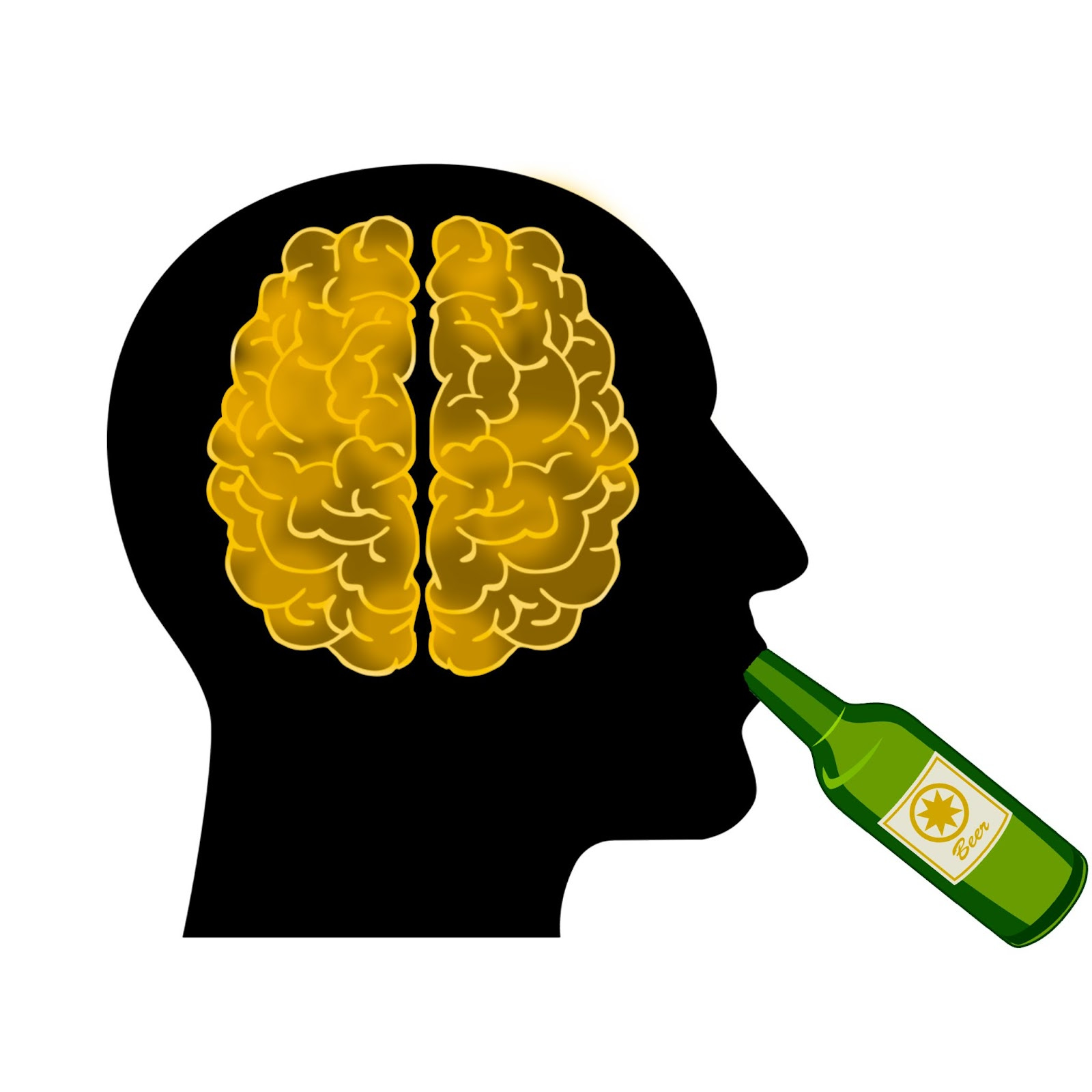
A Single Dose of Alcohol Produces Significant and Lasting Changes in Brain Morphology:
Although the dangers of binge drinking are well-known, many would argue that drinking in moderation is a mostly harmless way to unwind. However, a recent study out of Heidelberg University suggests that a little alcohol can go a long way in terms of altering the structure of the brain, perhaps permanently. Getting drunk even a single time significantly impacts the morphology of the brain, and produces functional changes that may contribute dangerously to addiction.
🧠 Article: “Single-dose ethanol intoxication causes acute and lasting neuronal changes in the brain” (August 26) - J. Knabbe et al.,(2022) Single-dose ethanol intoxication causes acute and lasting neuronal changes in the brain. Proc. Natl. Acad. Sci. U.S.A. 119, e2122477119.
🧠 Introduction and Methods:
Previous research has shown that even a single instance of alcohol intoxication at an early age greatly increases one’s risk for abusing alcohol later in life. It is thought that this phenomenon may involve the hippocampus forming pleasurable memories around alcohol, and although studies have tracked the morphological changes that the hippocampus undergoes during chronic alcohol exposure, there is little literature regarding the impact of a single dose. This study attempted to uncover any lasting morphological changes in the mouse brain that arise after just a single exposure to alcohol.
The team first looked for changes in which synaptic proteins were expressed in the alcohol-dosed mice. Since the synapse is fundamental in neural plasticity, information storage, and cellular communication, any changes to their structure would be significant to overall brain function. The researchers examined over 2,000 synaptic proteins in the hippocampus of young mice using quantitative mass spectroscopy to uncover differences in protein expression. They then used microscopy to reveal any visible changes in cell structure.
🧠 Results and Discussion:
The team found that a single dose of ethanol produced lasting changes in the abundance of 72 different synaptic proteins in the hippocampus, only 27 of which had previously been linked to alcohol. Notable among these 72 proteins were a group that affected the mitochondria of the synapse and two that filled important structural roles in the cell.
The identified mitochondrial proteins were expressed more abundantly after alcohol exposure, and made it so that mitochondria (responsible for cellular energy production) were more mobile between organelles and more susceptible to displacement. After running a brief experiment on fruit flies, the team found that this increased mitochondrial trafficking may affect the dopaminergic system in a way that associates alcohol with more rewarding memories.
The two structural proteins that had their concentrations altered, ankyrin-G and MAP6, affect the stability of dendritic spines and the morphology of the axon. Examining neurons under the microscope revealed that the mice who were exposed to a dose of alcohol experienced twice as much spine turnover as non-exposed mice, and that the initial segment of their axons was significantly shorter. The team also identified changes in protein expression that would directly alter the abundance of specific neurotransmitters, especially by shutting down GABA reuptake transporters.
All of this combines to paint a pretty drastic picture of the lasting neural changes that arise from a single exposure to alcohol. According to the research team, “the identified proteins linked acute alcohol intoxication to alterations in synaptic trans-mission/plasticity, mitochondrial function, mood, apoptosis, and neurodegenerative diseases.” Not only can one instance of intoxication lead to an alcohol dependence, it can have dangerous effects on the structure of the brain. Though consistent binge drinking is the most harmful way to consume alcohol, even sporadic drinking can be permanently destructive to our mental faculties.
Yale Scientists Uncover What Makes the Human Brain Unique:
Though humans are obviously unique in the animal kingdom for our ability to reason, understand, and think abstractly, the neural basis of our exceptionalism remains largely unknown. Scientists think the answer might lie in the dorsolateral prefrontal cortex (dlPFC), a brain region shared only by humans and our primate relatives. But is there something hidden in the dlPFC that is specific only to us, which separates the human brain from those of even our closest ancestors? Yale researchers may have some clues.
🧠Article: “Molecular and cellular evolution of the primate dorsolateral prefrontal cortex” (August 25, 2022) - Ma, S., Skarica, M., et al. (2022). Molecular and cellular evolution of the primate dorsolateral prefrontal cortex. Science, 377(6609), 999–1004. https://doi.org/10.1126/science.abo7257
🧠Introduction and Methods:
The dlPFC is partially responsible for the inter-region connectivity and higher-order cognitive abilities that separate primates from other orders of animals. This brain region is linked to many of the complex mental functions that we think of as “human,” including planning, inhibition, and abstract reasoning. Since this brain region may hold clues about why humans and other primates are so cognitively advanced compared to other organisms, the research team wanted to delve into the evolution of the dlPFC by examining over 600,000 transcriptomes (a full collection of the RNA molecules derived from a cell’s DNA) in marmosets, macaques, chimpanzees, and humans. The team used computational analysis to examine inter-species differences that may teach us about the development of this brain region and the factors that make the human brain unique.
🧠Results and Discussion:
The study monitored the RNA expression of neurons and glial cells in the dlPFC of each primate. Out of all the cells examined, just five were specific to particular species. The only cell type that was unique to humans was a category of microglial cell, integral in the brain’s immune system, that expressed the gene FOXP2. They also found that humans have an extra population of previously unknown dopaminergic interneurons in the prefrontal cortex. This additional helping of dopamine may be crucial in differentiating human thought and behavior from other primates.
In addition to satisfying curiosities about what makes the human brain special, the team hopes that this study will assist clinical researchers in developing human-specific treatments in the future. Though the RNA sequencing process ultimately showed far more similarities than differences between humans and our monkey ancestors, for the first time ever we know that at least one cell in the all-important dlPFC separates us from the pack.
If you missed our Neural Book Club post you can check it out here! We highly recommend Livewired by David Eagleman, so check it out with the link below.
Livewired: The Inside Story of the Ever-Changing Brain 👉 https://amzn.to/3PMPNDO
very interesting (about the alcohol effect on the brain) !
But I don't think it's chronic