
The Neural Newsletter 11/7-11/14
🧠 Last Week in Neuroscience

If you find value in our zero-cost information, you can support the newsletter by sharing it with a friend or by using the following links to get one of the curated neuroscience books we reviewed for our book club: Livewired by David Eagleman or The Body Keeps the Score by Bessel van der Kolk. Thanks for reading!
🧠Dopamine’s Role in Processing Aversive Stimuli
Dopamine is one of the brain’s most dynamic neurotransmitters, influencing a vast range of behaviors including learning and motivation. Many proposed functions of this powerful chemical remain controversial, with one line of debate questioning its role in processing aversive stimuli. Some researchers claim that dopamine is not involved in this process at all, while others argue it plays a crucial role in signaling aversive prediction error. Dopamine in the nucleus accumbens has proven pivotal in reward prediction error, or the signaling of a difference in expected versus actual reward to drive reinforcement learning. A team from the Royal Netherlands Academy of Arts and Sciences wanted to determine whether this brain region similarly utilized dopamine to signal prediction errors when it came to aversive stimuli. If not, they wanted to determine another model for dopamine’s role in aversive stimulus processing.
🧠Article: “Nucleus accumbens dopamine tracks aversive stimulus duration and prediction but not value or prediction error” (11/11/2022) Jessica N Goedhoop, Bastijn JG van den Boom, Rhiannon Robke, Felice Veen, Lizz Fellinger, Wouter van Elzelingen, Tara Arbab, Ingo Willuhn (2022) Nucleus accumbens dopamine tracks aversive stimulus duration and prediction but not value or prediction error eLife 11:e82711
🧠Introduction and Methods:
Dopamine’s main functions relate to motivation and learning; the molecule is responsible for generating motivational states that encourage reward-seeking and for facilitating reward-based learning. A crucial mechanism in this dopamine-driven learning process is reward prediction error, a process through which the difference between an expected reward and a received reward is encoded via a difference in dopamine release. When receiving a predicted reward dopamine neurons will not fire, but if the reward is unexpected they will fire rapidly and increase rates of learning.
Though dopamine’s relationship to rewards, or positive valence stimuli, is well studied, less work has been done with regard to aversive, negative valence stimuli. Many studies have produced seemingly contradictory results, but consistent findings suggest that dopaminergic activity changes in response to aversive stimuli and that this activity varies across regions of the striatum, a decision-making, motivation, and reward center, in a somewhat unpredictable fashion. To parse out these uncertainties, the team found it necessary to delineate the specific function of aversive dopamine fluctuations: did it encode an aversive prediction error (capturing the discrepancy between an expectation of an aversive stimulus versus the one received) or did it simply signal the presence of an aversive stimulus?
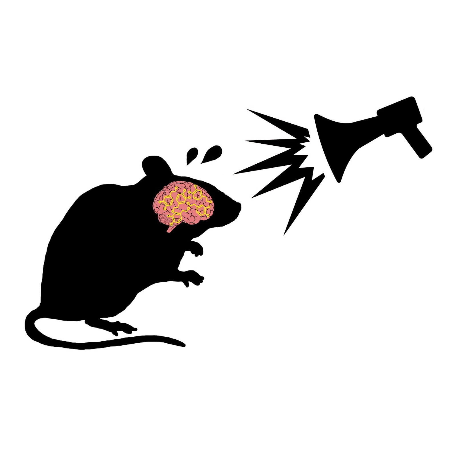
The researchers did this by varying the value of an aversive white noise stimulus in mice and by altering probability contingencies, conditioning states, and other contextual elements of the experiment. They used fast-scan cyclic voltammetry to track dopamine in the nucleus accumbens, a known hotspot in reward prediction error as well as aversion avoidance motivation. If dopamine plays a role in aversive prediction error as has been hypothesized, the team’s measurements would produce some chemical pattern resembling reward prediction error in the nucleus accumbens.
🧠Results and Conclusions:
Initially, the team used approach-avoidance tests and operant conditioning to establish that white noise was indeed an aversive stimulus for mice, and that its aversiveness scaled with volume intensity based on behavioral observations. Accordingly, they found that the nucleus accumbens dopamine response to aversive stimuli was opposite in directionality compared to its reward response. Where rewards produce an increase in measurable dopamine, aversive stimuli caused a decrease.
They also found that in conditioning tasks where an auditory cue precedes the white noise, the conditioned auditory cue quickly produces a similar dopamine-depressing response. This indicates that dopamine is affected by the prediction of an aversive stimulus, not just the experience of the stimulus itself.
Interestingly, the team found that dopamine reductions did not change as the white noise stimulus became more intense/aversive, nor did they change with the introduction of intermittent rewards or altered stimulus probabilities. Instead, they noted a predictably slow and steady ramping-down of dopamine as the negative stimulus was predicted and experienced, followed by an equal ramping-up effect after the stimulus ceased. The dopamine down-ramp does not appear to plateau, changing its slope only when the aversive stimulus is removed. The slope of the ramping-up response could be altered by the unpredictable presentation of the aversive white noise, but not by making it unpredictably more or less intense. This indicates that, contrary to the team’s hypothesis, dopamine in the nucleus accumbens does not seem to encode prediction error when it comes to aversive stimuli. Instead, it seems to track the prediction and duration of an aversive event without encoding any information about the level of aversiveness. If dopamine does dictate aversion prediction error, it appears that this process occurs in a less obvious region of the brain.
“Dopamine tracks both positive and negative valence in their temporal aspects and prediction, but quantitatively speaking, the exact value and error is only encoded for rewards, in the upward direction of [nucleus accumbens] dopamine concentration. This implies that aversive value and aversive prediction errors are encoded in other brain regions.”
-Goedhoop et al.
Autism Brain Changes are Surprisingly Wide-reaching
Autism spectrum disorder, like many neuropsychiatric disorders, has been famously difficult to link to structural brain changes. However, recent research has begun to dig up potential mechanisms for ASD, noting discrepancies in gene expression that may contribute to autism symptoms. Little is known about the generality of these epigenetic changes, and researchers are unsure whether they impact the whole cortex or if they’re limited to the association areas controlling social behavior and higher-order processing. A team from the University of Pennsylvania sought to better understand the molecular basis of autism by analyzing post-mortem brain tissue samples of autistic individuals. Their work uncovered crucial findings about the mechanisms and scope of autism-related brain changes, potentially laying the groundwork for clinical interventions down the line.
🧠Article: “Broad transcriptomic dysregulation occurs across the cerebral cortex in ASD” (11/7/22) - Gandal, M.J., Haney, J.R., Wamsley, B. et al. Broad transcriptomic dysregulation occurs across the cerebral cortex in ASD. Nature (2022). https://doi.org/10.1038/s41586-022-05377-7
🧠Introduction and Methods:
Like many other neurodevelopmental disorders, autism is strongly rooted in genetics. The complexity of ASD’s genetic component has proven baffling to researchers, with hundred of risk genes interacting to form an indecipherable puzzle. Despite numerous contradictions across the literature, most studies identify consistent disturbances in RNA and gene expression in the frontal and temporal cortex.
UPenn researchers felt that a better understanding of these molecular patterns would produce a more refined neurobiological picture of autism spectrum disorder. To that end, they analyzed changes in RNA and gene expression in post-mortem brain tissue samples from 49 autistic individuals. They also included a neurotypical control of 54 population-matched subjects to examine structural differences. In total, the researchers studied 725 brain samples from 11 cortical areas. They took RNA profiles from each sample to determine which neural proteins were expressed, and used these findings to model brain differences between autistic and neurotypical subjects. They mainly wanted to determine whether molecular discrepancies were localized to specific cortical regions, or if the changes were more widespread.
🧠Results and Conclusions:
The team’s findings significantly challenge the prevailing belief that molecular differences in ASD are mainly confined to the frontal and temporal lobes. Extensive epigenetic and transcriptomic analysis revealed changes across the cerebral cortex, affecting not just higher-order association areas but also more fundamental sensory regions of the brain. In fact, the most prevalent alterations in gene expression could be found in the primary visual cortex. Although these neurobiological changes have never been previously identified, they are consistent with the vast differences in sensory processing that characterize autism.
Many crucial neural processes were impacted in autistic individuals, including a downregulation in blood-brain-barrier transport and an upregulation in astrocyte number and reactivity. This is part of a cortex-wide upregulation in the neural-immune response. There also appears to be a widespread reduction in synaptic plasticity and neural circuit function, which may be linked to an overall disruption in cortical architecture. Specifically, the GeneM5 co-expression module, which represents genes that affect synaptic plasticity, is down-regulated across the cortex and may play a causal role in ASD.
As it stands, this study has revealed that the molecular changes in ASD are far more delocalized and pervasive than previous models suggest. Further experimental studies are needed to expand on these rich insights, hopefully drawing us closer to effective treatments and preventions for autism.
“The findings presented here substantially refine our understanding of ASD molecular pathology beyond the previously established ‘downregulated neuron’ and ‘upregulated glia/immune’ functional categories observed in frontal and temporal lobes. We identify gene and transcript expression changes in ASD that occur across the cerebral cortex, affecting many neural cell types and specific biological processes, extending beyond higher-order association areas to include primary sensory areas.”
- Gandal et al.
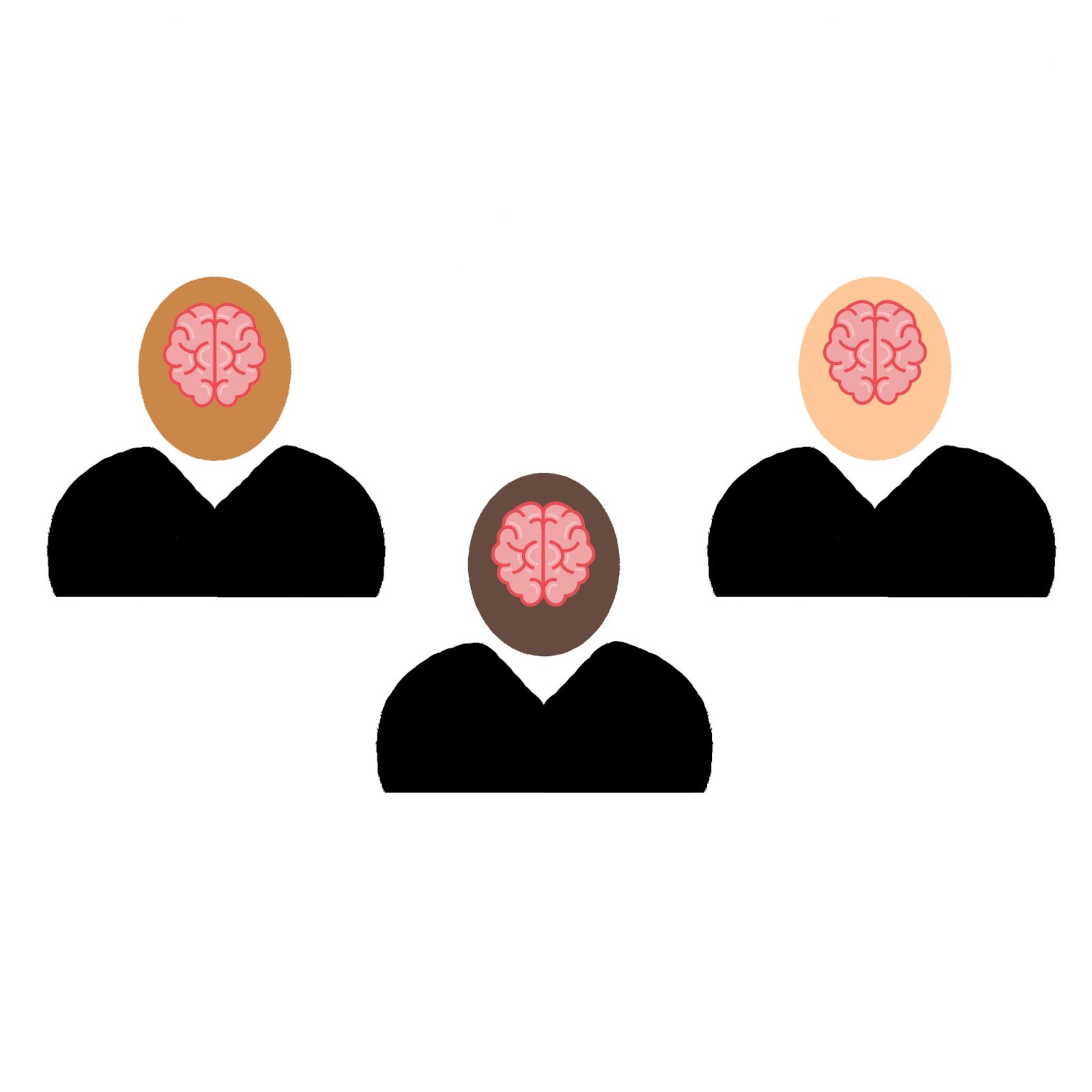
🧠Differential Brain Aging Among Racially Diverse Americans
Previous neuroimaging work has identified a disparity in elderly brain health along racial and ethnic lines, presumably due to societal and economic disadvantages. Little has been done to study these differences earlier in life, leaving many questions about the nature and extent of racial brain differences due to socioeconomic standing. A research team from Columbia University sought to address this by using Alzheimer's-related neuroimaging to track trends in neurological racial disparities from middle age to late life.
🧠Article: “Brain Aging Among Racially and Ethnically Diverse Middle-Aged and Older Adults” (11/14/2022) - Turney IC, Lao PJ, Rentería MA, et al. Brain Aging Among Racially and Ethnically Diverse Middle-Aged and Older Adults. JAMA Neurol. Published online November 14, 2022.
🧠Introduction and Methods:
It’s well established that racial minority groups exhibit relatively diminished levels of brain health in old age compared to White American populations. The “weathering hypothesis” suggests that members of ethnic minority groups might experience accelerated physiological wear due to stress, material disadvantage, and lack of social support. In a neuroscience context, this has been proposed to contribute to differential brain aging and increased neurodegenerative disease in Black and Latino communities. The research team chose to examine midlife and late-life populations across racial groups to look for differential patterns in brain aging, as determined by cortical thickness measurements and cerebrovascular disease( based on white matter hyperintensity volume). Both of these markers can be measured using magnetic resonance imaging (MRI), and both have been implicated in the onset and progression of Alzheimer’s Disease.
The study included participants from the Washington Heights–Inwood Columbia Aging Project and the Offspring Study of Racial and Ethnic Disparities in Alzheimer Disease. The team examined data from 497 midlife participants [mean age 55] and 970 late-life participants [mean age 75], most of whom were Black or Latino. These studies examined single communities, so environmental confounds were limited. They performed statistical comparisons to uncover insights about differences in cortical thickness and white matter hyperintensity based on these populations, resulting in insights about brain aging variability across racial groups.
🧠Results and Conclusions:
The team’s statistical analyses exposed a number of important patterns between groups. They found that racial differences in cerebrovascular disease markers were present in midlife as well as late life, while cortical thickness diverged only in late life. Brain measures between Black and Latino groups did not differ significantly, but both groups showed disparities compared to White populations. Black-White differences were consistently the largest across measurement categories. The racial disparity in white matter hyperintensity was greater in midlife than in late-life, which may stem from differences in how likely these populations are to survive into the late-life age group.
The rate of cortical thickness and white matter hyperintensity reduction was significantly greater in old age than in mid-life among White and Latino groups, while it remained mostly stable for Black participants. This may indicate that for Black populations, the inflection from a biologically young brain to a biologically old brain occurs earlier than in White or Latino groups. This suggests that the socioeconomic and psychosocial stress that Black Americans experience leads to measurably accelerated brain aging compared to White or Latino Americans. Now that we have an added body of evidence for aging patterns and neural changes that support the weathering hypothesis, researchers must examine the mechanisms that cause this disturbing discrepancy so that we may begin to address it.
“The WHICAP and Offspring studies include participants who have been historically excluded from studies of brain aging and Alzheimer’s Disease. Data from both community-based studies provide a unique opportunity to examine the role of race and ethnicity across a wide age range of participants (age 25-98 years), who also have health status, economic, and educational backgrounds that are representative of their communities. This study shows that race and ethnicity disparities observed at older ages are apparent in midlife, suggesting accelerated brain aging in Black participants.”
Turney et al.
Enlightening. Thanks for a great summary.
Very interesting indeed! Thank you, Frederick