

If you find value in our zero-cost information, you can support the newsletter by sharing it with a friend or by using the following links to get one of the curated neuroscience books we reviewed for our book club: Livewired by David Eagleman or The Body Keeps the Score by Bessel van der Kolk. Thanks for reading!
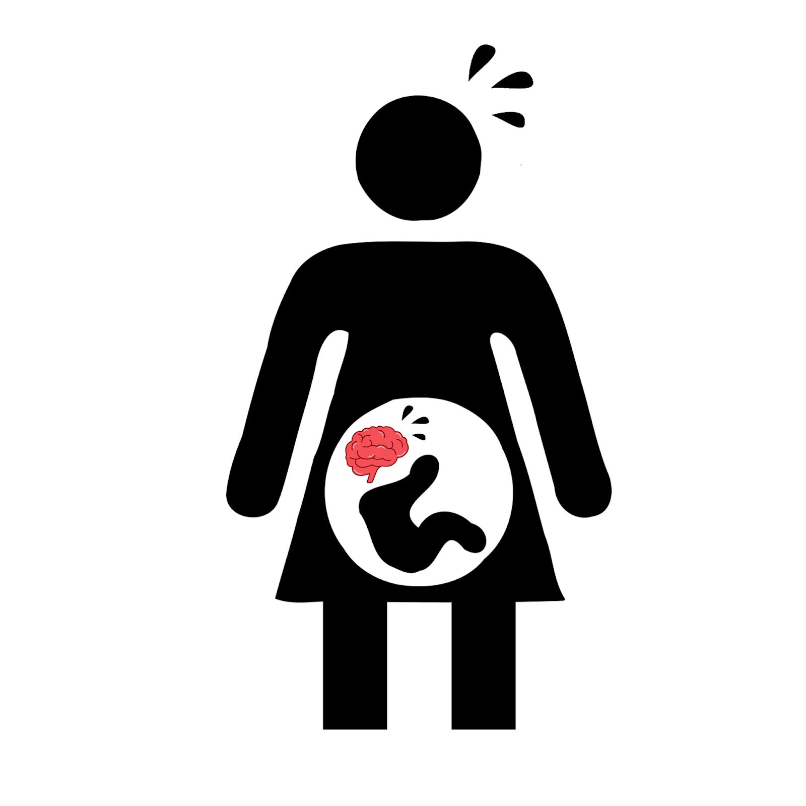
🧠 Social Disadvantage and Psychosocial Stress Harm the Prenatal Brain
Early life adversity and psychosocial stress are known to disrupt the physical development of the brain, perpetuating cycles of social inequity across generations. However, it’s unclear exactly when these factors begin to affect neural development, and whether there are critical windows in which their effects are most pronounced. A recent neuroimaging study out of Washington University shows that these issues must be addressed early, as an infant’s white matter microstructure is measurably affected by maternal stress even before the child is born. The team identifies a critical prenatal window during which the child’s fronto-limbic pathways are especially sensitive to adverse environmental conditions, and cites the external factors which contribute most potently to aberrant development.
🧠Article: “Prenatal exposure to maternal social disadvantage and psychosocial stress and neonatal white matter connectivity at birth” (10/03/2022) - Fastenrath, M., Spalek, K., Coynel, D., Loos, E., Milnik, A., Egli, T., Schicktanz, N., Geissmann, L., Roozendaal, B., Papassotiropoulos, A., & de Quervain, D. J.-F. (2022). Human cerebellum and corticocerebellar connections involved in emotional memory enhancement. Proceedings of the National Academy of Sciences, 119(41). https://doi.org/10.1073/pnas.2204900119
🧠Introduction and Methods:
Stressors caused by social disadvantage have been shown to produce damaging and permanent effects on the brain in critical periods during early childhood. Thus far, scientists have struggled to identify the precise time windows during which the child is most sensitive to this neurological damage, but they have demonstrated that disadvantaged children tend to show a size reduction in the amygdala, hippocampus, and prefrontal cortex. Since this effect is so disturbing, Washington University researchers wanted to learn just how early social disadvantage begins to stress the developing brain. They chose to study the development of white matter (nerve fibers as opposed to cell bodies) while a child is still in utero, and to gauge how different environmental factors impact structure and connectivity.
To achieve this, the team performed a diffusion MRI (dMRI) procedure on 289 healthy newborn babies. They collected a range of variables to measure the mother’s social disadvantage and psychosocial stress, including racial discrimination, income-to-needs ratio, depression, nutritional access, neighborhood safety, health insurance, education, and more. They focussed their imaging on the cingulum bundle, uncinate, and fornix, which are key components of the fronto-limbic pathway that connects the emotional limbic system to the planning frontal cortex. They also included the corpus callosum and corticospinal tracts as controls, expecting to see impaired brain development in the fronto-limbic system but not these unconnected regions. They also used statistical techniques to try to isolate the effects of maternal psychosocial stress from social disadvantage while eliminating confounding factors like drug exposure.
🧠Results and Conclusions:
The team took measures of mean diffusivity (MD) and fractional anisotropy (FA) to measure brain connectivity in the regions of interest. They found that prenatal exposure to social disadvantage caused major alternations in fronto-limbic brain connectivity at birth, varying between increased and decreased MD and FA measures across different parts of the uncinate, cingulum bundle, and fornix. This inconsistency may reflect aberrant and unpredictable patterns in nerve branching and pruning, as well as premature myelination and glial differentiation. If this premature hypermaturation hypothesis is true, it holds with previous research that demonstrates an overactive adrenal response in people who experienced accelerated amygdala-frontal connectivity due to early life adversity.
The study specifically identified higher levels of FA in the cingulum bundle and uncinate in socially disadvantaged newborns, which is predictive of autism and socioemotional issues. Social disadvantage had less of an impact on the control brain regions than the fronto-limbic regions of interest, but still had a minor effect on the corticospinal tract which means that the damage caused by prenatal adversity may be more wide-ranging than the researchers anticipated.
Interestingly, psychosocial stress did not have as clear an impact on white matter development when separated from markers of social disadvantage, especially among the newborns of lowest socioeconomic status. However, newborns in a slightly higher socioeconomic group saw alterations to their left anterior cingulum bundle when their mother was more stressed. The team found that of all the factors measured, income-to-needs ratio was the most predictive of gray matter changes in the neonates.
Ultimately, the study found that social disadvantage and psychosocial stress in utero produce significant changes in fronto-limbic white matter microstructure, possibly contributing to socioemotional issues such as autism. This means that early-life adversity begins to affect brain development even before birth, and is especially potent amongst children of the lowest socioeconomic status. These findings should compel us to extend further help to disadvantaged families, or else subject children to a continuing cycle of unfair circumstances for generations to come.
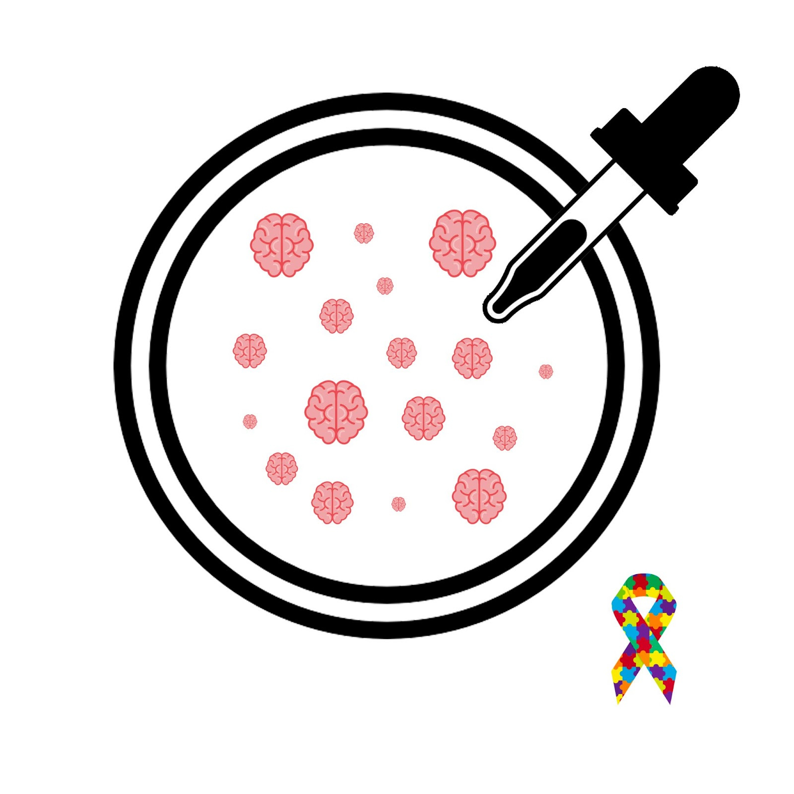
🧠Stem Cell Organoids Mimic Human Forebrain Development
The telencephalon (or forebrain) comprises 85% of the human brain, including all of its most evolutionary advanced and unique structures. Neuroscientists have had a hard time studying the telencepahlon’s complex development, but a fascinating new technique allowed a University of Utah team to examine this structure’s early organization under both normal and pathological conditions. Using pluripotent STEM cells, the researchers created seed-sized, brain-like structures called organoids which can be made to emulate specific brain regions. After showing that these organoids can help simulate normal telencephalon development, they introduced changes to the autism-related SHANK3 gene to show that this method can also be used to study aberrant neural development.
🧠Article: “Modeling human telencephalic development and autism-associated SHANK3 deficiency using organoids generated from single neural rosettes” (10/06/2022) - Wang, Y., Chiola, S., Yang, G. et al. Modeling human telencephalic development and autism-associated SHANK3 deficiency using organoids generated from single neural rosettes. Nat Commun 13, 5688 (2022). https://doi.org/10.1038/s41467-022-33364-z
🧠Introduction and Methods:
Since the telencephalon comprises the most complex parts of the brain, it’s linked to many of the disorders and behaviors that are unique among humans. Despite this intrigue, the region has always been difficult to study and its development has remained mysterious to scientists. Researchers have used RNA sequencing to map the gene expression of telencephalic cells, but understanding the molecular mechanisms that allowed these cells to differentiate required new techniques.
The University of Utah team wanted to test whether telencephalon-specific organoids could effectively model the neural development of this elusive brain region. In other words, the team used self-organizing human stem cells to create tiny organic structures that mimic the human forebrain both in development and activity. After showing that these lab-grown organoids could reproducibly emulate healthy telencephalon organization, the team implemented a deletion of the SHANK3 gene, commonly implicated in autism, to see whether their method could be a useful tool in studying neurodevelopmental disorders. The organoids were allowed to organize themselves without interference for a month, at which time they were subjected to single-cell RNA sequencing and statistical techniques to measure their similarity to the human telencephalon and to tests the consistency of the protocol.
🧠Results and Conclusions:
The study showed that stem cell-derived organoids can successfully model the complex early development of the human telencephalon, complete with functional neurons and synapses characteristic of this brain region. This process may be the most naturalistic way to model brain development, as it mimics the starting conditions that the human brain experiences in vivo.
They showed that a single cluster of telencephalic neural progenitor cells could produce a structure composed of a diverse array of neurons and glia that we would expect to find in the human forebrain. Interestingly, these organoids also contained the ependymal cells and periendothelial cells which help form brain barriers, even though these cells have not been identified in previous organoid studies. They showed that both inhibitory and excitatory synapses developed quickly in the telencephalic organoids, and were able to measure neural oscillations between 4 and 100 Hz to show that theses cells were functioning.
After showing that their method could credibly simulate early telencephalon development, they applied their technique to neurological disorders by growing organoids with the damaging SHANK3 deletion. Surprisingly, they found that this mutation did not significantly impact the cell identities in the organoids. However, the deletion did cause a reduction in excitatory synapses within the telencephalic tissue. One possible mechanism for this difference is the team’s finding that SHANK3-deficient organoids expressed fewer protocadherin clusters, which are protein complexes that affect synaptogenesis and proper brain wiring. However, while correlation has been established, the mechanistic link between SHANK3 deletion and protocadherin deficiency deserves further study.
Ultimately, this study introduces an exciting new method for researching brain development under normal or pathological conditions. By using organic organoids that can mimic specific brain regions, the researchers uncovered insights about the development and composition of the telencephalon as well as the neurobiological underpinnings of the autism-related SHANK3 deletion. In doing this, the team has presented a compelling proof of concept for a method that promises to produce even more impactful findings in the future.
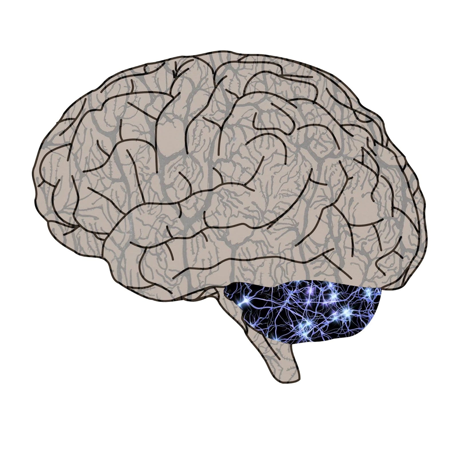
🧠The Cerebellum’s Surprising Role in Emotional Memory Storage
The cerebellum is a primitive brain structure, historically known for its role in balance and movement. While this was once thought to be this region's only function, modern work has revealed that the cerebellum communicates with the cortex to play a surprising role in specific higher-order tasks. Most recently, researchers out of the University of Basel used fMRI to show that this ancient structure interacts with the limbic system to handle emotionally weighted memories. Understanding the cerebellum and this pathway as a whole may help us better understand trauma moving forward, as well as numerous other psychiatric disorders which involve memory and emotion.
🧠Article: “Human cerebellum and corticocerebellar connections involved in emotional memory enhancement” (10/03/2022) - Fastenrath, M., Spalek, K., Coynel, D., Loos, E., Milnik, A., Egli, T., Schicktanz, N., Geissmann, L., Roozendaal, B., Papassotiropoulos, A., & de Quervain, D. J.-F. (2022). Human cerebellum and corticocerebellar connections involved in emotional memory enhancement. Proceedings of the National Academy of Sciences, 119(41). https://doi.org/10.1073/pnas.2204900119
🧠Introduction and Methods:
Humans have evolved to encode emotional events more intensely in our memories, allowing us to modify our future behavior in response to highly aversive or rewarding outcomes. Although this is necessary for our survival, it also allows traumatic baggage and fear-related disorders to take hold. This process is mostly due to increased connectivity between the amygdala and hippocampus when an emotional stimulus elicits a noradrenergic response, but recent work has shown that this process is aided by multiple other brain regions. The cerebellum, a primitive hindbrain structure that means “little brain” in Latin, has been implicated in unconscious fear conditioning. However, it is yet unclear whether it helps encode emotionally salient episodic memories, though this process seems to overlap with fear conditioning pathways.
The Basel University team wanted to understand the cerebellum’s role in emotional memory through an fMRI task. They showed a sample of 1418 participants images that were either positive, negative, or neutral in their emotional content. They then asked the participants to recall these images, and defined regions of interest based on which parts of the brain produced the greatest fMRI response when remembering emotional pictures. They found responses in both the cerebellum and the cerebrum, and used dynamic causal modeling to map connectivity between the regions of interest. Using this framework they could examine whether these connections became stronger during emotional recollection and whether the cerebellum causally drives a response in the cerebrum as this occurs.
🧠Results and Conclusions:
As expected, the team’s neuroimaging procedure revealed numerous cerebral clusters that responded to emotional episodic memory, in regions that include the frontal cortex, amygdala, hippocampus, and thalamus. Notably, they also found evidence that the cerebellum was crucially involved as well. The team found a clear cluster of activation in the cerebellar vermis, the strip of tissue which separates the two hemispheres of the cerebellum. This region is also significantly activated in fear conditioning, and has projections to emotional and memory-related brain centers like the amygdala and hippocampus.
They also found that 11 connections between the cerebellum and the cerebrum increased in strength during emotional memory retrieval, suggesting an integrated network between these brain areas. Dynamic causal modeling suggests that the cerebellar vermis influences the limbic system, as well as brain regions involved in valuation and motor learning.
This information may have clinical implications, as the midline cerebellum can now be considered a major region of interest when it comes to emotional memory disorders like PTSD. It also supports the idea that emotional deficits in disorders like autism may be linked to cerebellar dysfunction. By delivering input to the amygdala and hippocampus, the cerebellum emerges as a major contributor to the emotional enhancement of memory and should be studied further to better comprehend an important array of psychiatric disorders.