
The Neural Newsletter 10/10 - 10/17
🧠 Last Week in Neuroscience

If you find value in our zero-cost information, you can support the newsletter by sharing it with a friend or by using the following links to get one of the curated neuroscience books we reviewed for our book club: Livewired by David Eagleman or The Body Keeps the Score by Bessel van der Kolk. Thanks for reading!
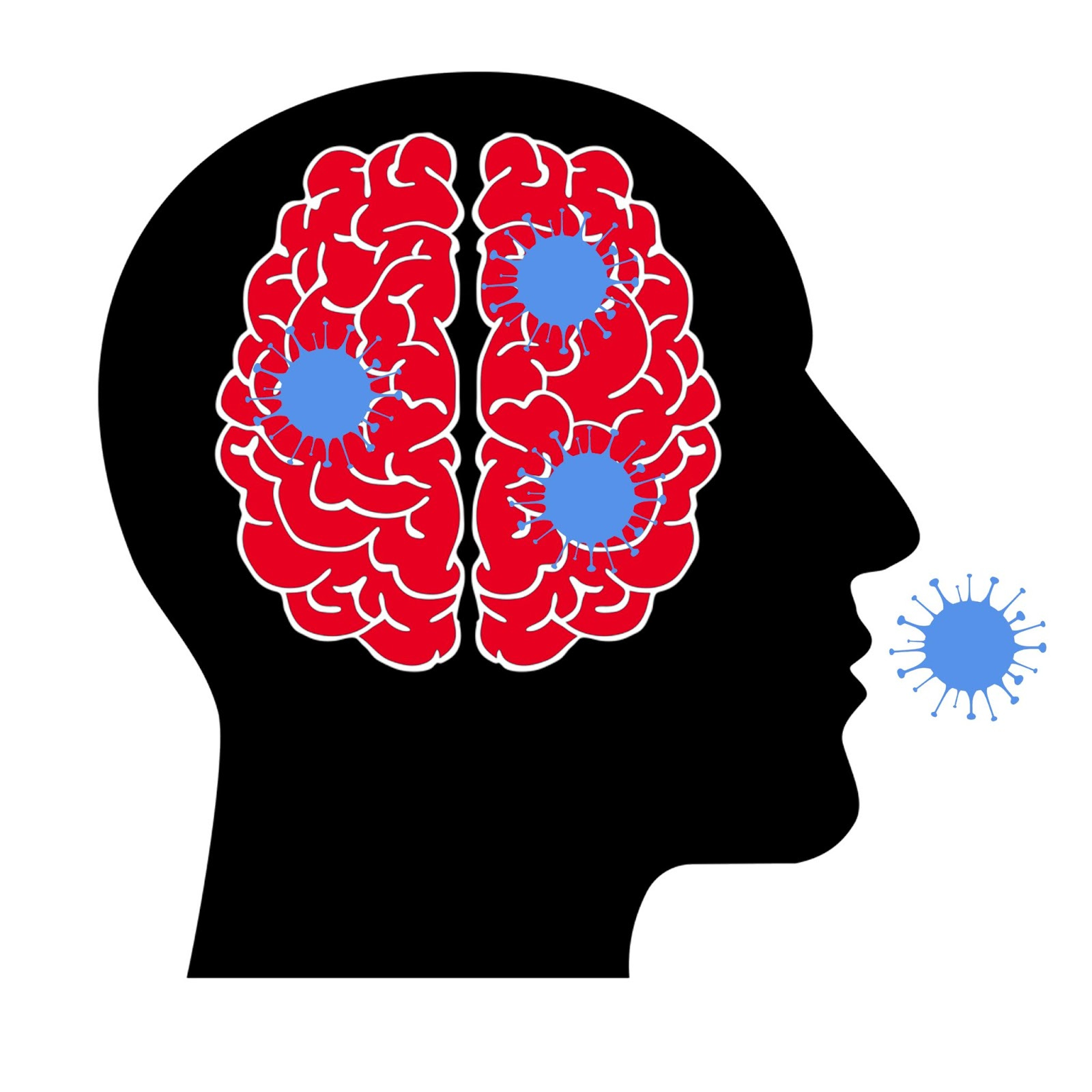
🧠 Neurocovid Infects and Inflames Brain Cells
COVID-19’s disturbing neurological effects have garnered major media attention, but our scientific understanding of these symptoms lags behind. A UC Davis team wanted to determine whether these complications were caused by the virus directly invading the brain, or if they came from a broader inflammatory response that affected the nervous system indirectly. The study develops a cellular and molecular understanding of COVID's impact on the brain, which may help relieve some of the virus' damaging neural side effects in the future.
🧠 Article: “SARS-CoV-2 infects neurons and induces neuroinflammation in a non-human primate model of COVID-19” (10/13/22) - Danielle Beckman, Alyssa Bonillas, Giovanne B. Diniz, Sean Ott, Jamin W. Roh, Sonny R. Elizaldi, Brian A. Schmidt, Rebecca L. Sammak, Koen K.A. Van Rompay, Smita S. Iyer, John H. Morrison,
SARS-CoV-2 infects neurons and induces neuroinflammation in a non-human primate model of COVID-19, Cell Reports, 2022, 111573, ISSN 2211-1247, https://doi.org/10.1016/j.celrep.2022.111573.
🧠 Introduction and Methods:
COVID-19, a single-strand RNA coronavirus, has dominated the public consciousness for the last two years as it infected almost 100 million people in America alone. Though its respiratory symptoms are the most apparent, the virus also produces neurological symptoms and gray-matter reductions in up to 80% of hospitalized patients.
Post-mortem brain samples have failed to determine whether the virus invades the brain directly, or if the neural symptoms are indirect byproducts of some other viral behavior. To uncover the crucial mechanism for these symptoms, researchers at UC Davis’ California National Primate Research Center used high-resolution 3D confocal microscopy to study the brains of rhesus macaques infected with COVID-19. They examined one cohort of young, healthy monkeys and another cohort of old, diabetic monkeys to see how the virus interacted with the brain across health demographics. They euthanized the monkeys 7 days after infection and used labeling techniques to see if a viral load accumulated in the brain and to record any signs of neurological damage.
🧠 Results and Conclusions:
Even though the monkeys’ COVID symptoms were usually still mild when their brains were analyzed a week after being infected, an immunohistochemistry analysis showed that the virus made a large-scale entrance into the central nervous system. COVID markers were mostly localized to the olfactory cortex, and were more widespread in older monkeys than younger ones. Both glial cells and neurons showed evidence of containing viral proteins, but the concentration was significantly higher in neurons.
After confirming that the virus entered the brain and occupied the olfactory cortex, the researchers wanted to understand the neuroinflammatory response which can damage cells. They found a much greater concentration of astrocyte and microglia cells in infected subjects, indicating a robust inflammatory immune response. They found that this inflammation was associated with myelin degradation and synaptic pruning, which may explain some of the neurological deficits that accompany COVID. Once again, the findings were more severe in older populations than young ones.
COVID-19 spike protein is known to have a binding site in ACE2, a protein that is crucial in the cellular entry process. They found that ACE2 production was highly downregulated in the brain areas with the highest viral load, and that the local vasculature of infected areas seemed to be disrupted. This implies a structural disturbance of the blood-brain barrier, meaning that foreign cells can enter the brain and inflict potential damage.
The study only examined the brain 7 days post-infection, so it’s possible that other mechanisms affect the brain as the disease develops. Still, knowing that the virus infects the brain directly and understanding the damage caused by neuroinflamation may help guide clinical interventions and further research moving forward.
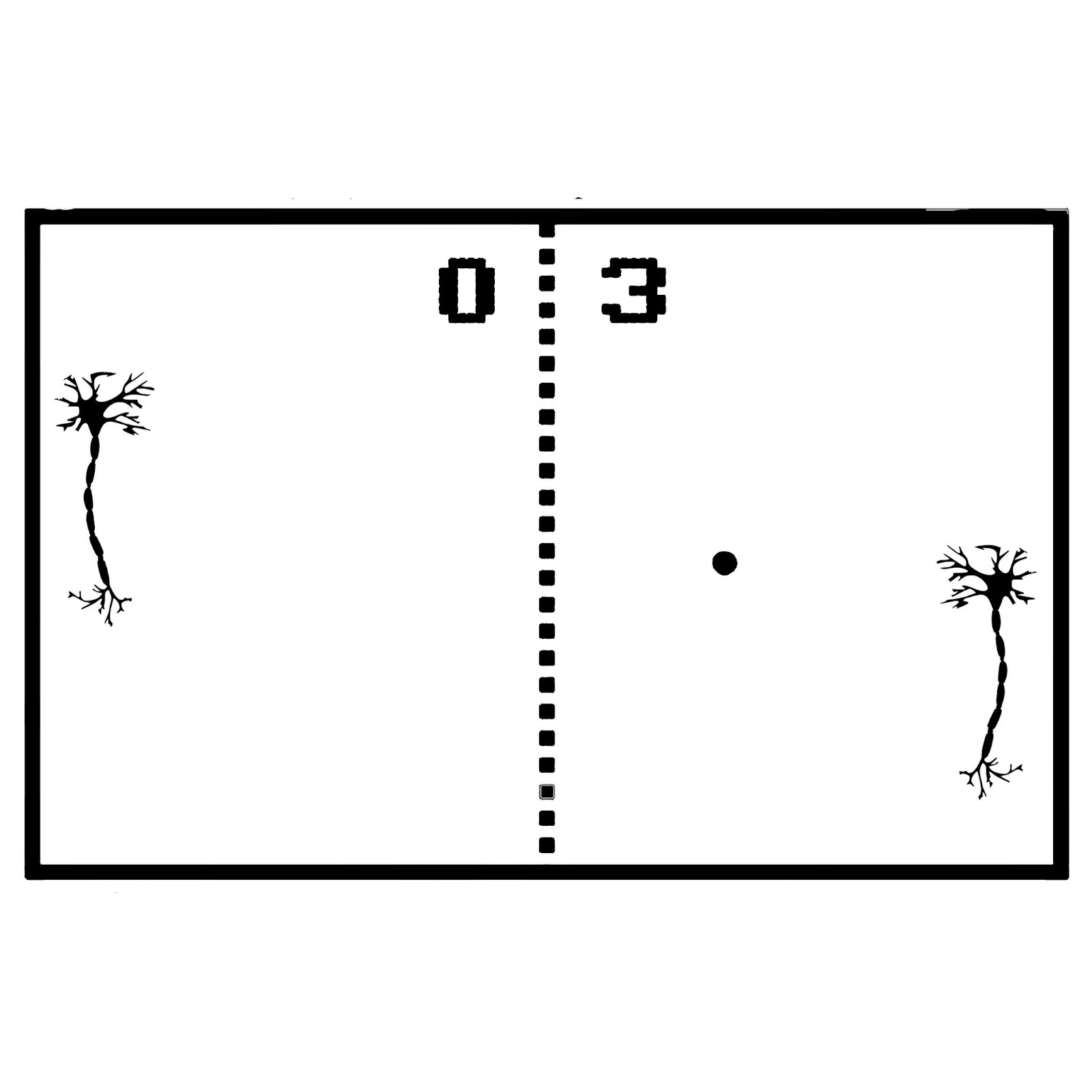
🧠 Gaming Neurons
Growing synthetic intelligence sounds like the stuff of science fiction, but it may not be as far off as it seems. Australian researchers have created a system called DishBrain that uses an electrode array to give live brain cells control over video game worlds. The experimenters allowed a dish of neurons to play the arcade game Pong, and found that the cells were able to self-organize to perform better at the game when provided with feedback. The team argues that these neural clusters meet the standard definition of sentience, and that studying their goal-oriented behavior at this basic level can build into a more comprehensive understanding of biological intelligence.
🧠 Article: “In vitro neurons learn and exhibit sentience when embodied in a simulated game-world” (10/12/22) - Brett J. Kagan, Andy C. Kitchen, Nhi T. Tran, Forough Habibollahi, Moein Khajehnejad, Bradyn J. Parker, Anjali Bhat, Ben Rollo, Adeel Razi, Karl J. Friston, In vitro neurons learn and exhibit sentience when embodied in a simulated game-world, Neuron, 2022, ISSN 0896-6273, https://doi.org/10.1016/j.neuron.2022.09.001.
🧠 Introduction and Methods:
Recent trends in neuro- and computer science have aimed towards hardware that mimics the biological functions of the brain. In spite of massive effort, no artificial solution has been able to handle the levels of computational complexity that our neurons manage with ease. Though countless studies have tried to model the computational underpinning of neural networks in the living brain, few have examined the computing power of neurons in vitro, outside of their biological environment.
Researchers from Cortical Labs in Melbourne addressed this challenge through a system they coined DishBrain. DishBrain consisted of stem cell-derived mouse and human cortical neurons grown on a high-density multielectrode array that records electrical signals. The team translated this neuronal firing data into paddle movements in the classic Pong video game, and gave the cells real-time electrical feedback based on their performance. In doing so, they hoped to create the first true example of synthetic biological intelligence, a biology-based computational device that can learn outside of its natural environment. Understanding how biological neural networks learn and organize outside of the brain could generate a flood of interdisciplinary innovations, ranging from drug-testing alternatives to machine learning developments to the manufacture of hybrid “silico-biological” computer hardware.
In their hopes of producing a sentient synthetic biological intelligence, the team had to address two requirements. Firstly, the agent must learn how the environment impacts its internal states through perception, and how its internal states can affect the environment through action. Secondly, it must be able to use its sensory state to predict the external outcome of a behavior, and decide when to choose certain behaviors based on this information. To address this first issue of learning through perception and action, the team created a feedback system based on Pong outcomes that induced neuroplasticity through electrical stimulation. To address the second issue of goal-directed decision making, the team modeled their feedback system based on a theory of intelligence called free energy minimization. Based on this framework, the DishBrain neurons were ascribed “beliefs” which updated to minimize the gap between the predicted outcome of an action and the observed outcome/sensation. If a neuron acted incorrectly, the team could give it a random, unpredictable electrical response (akin to a shocking outcome for a human behavior) which incentivizes the cell to avoid that behavior in the future. When they act correctly, in a way that results in the paddle making contact with the ball, all cells would instead receive a predictable stimulus of 100 Hz.
🧠 Results and Conclusions:
The team allowed their neurons to grow and make connections for over three months on the multielectrode array, forming a dense network of interconnected cells. Using their feedback system based on Pong outcomes, the team provided the first-ever demonstration of goal-directed learning in in vitro neurons. Within minutes, the neurons adapted their firing patterns to result in a greater percentage of paddle contact than they started with. This may be the first synthetic biologically intelligent system to model learning in real time.
The team found that the amount and diversity of outcome-based feedback they provided directly correlated with increased performance from the neurons. The more unpredictable the outcome stimulus, the greater the learning effect as the neural network learns to avoid uncertainty. Interestingly, the study also showed that human neurons outperformed mouse neurons at the task, providing empirical evidence that human neural networks are fundamentally superior at processing information even outside of the brain. The experiment also showed that dynamic self-organization of neurons can occur at the cellular level, removed from any higher-order input, as long as appropriate feedback is present. Importantly, the learning that these neurons gathered during an individual session seemed to reset back to baseline soon after the trial ended. The team expects that this is because the cortical neurons used were not specialized for long-term memory.
In the future, the team hopes that technoligcal advancements will afford greater precision to similar experiments. The researchers were unable to discriminatively target the dendrites or the soma of the cells, which limited the finesse with which they could induce neuroplasticity. If continue to progress towards harnessing the natural learning behaviors of brain cells, we may be able to revolutionize standard computational tools by endowing our electronic devices with biological intelligence.
🧠 How Goals Affect Our Senses
Research has shown that our internal motivational states influence our ability to process sensory information when performing a task. When we are in a more motivated state, we can learn and perceive information more effectively to reach a desired goal. However, this is only true to a point, as excessive levels of motivation can create pressure that distracts from the task and hinders performance. University of Geneva researchers wanted to examine the roots of this precarious system by having mice with varying levels of thirst motivation perform tasks for a water reward. The team uncovered important behavioral and mechanistic discoveries which explain how the brain gives rise to certain behavioral outcomes depending on an organism’s motivational state.
🧠 Article: “Cortical sensory processing across motivational states during goal-directed behavior” (10/13/22) - Giulio Matteucci, Maëlle Guyoton, Johannes M. Mayrhofer, Matthieu Auffret, Georgios Foustoukos, Carl C.H. Petersen, Sami El-Boustani, Cortical sensory processing across motivational states during goal-directed behavior, Neuron, 2022, ISSN 0896-6273, https://doi.org/10.1016/j.neuron.2022.09.032.
🧠 Introduction and Methods:
Sensorimotor transformations form the basis of perceptual decision-making. In other words, we respond to decisions by taking in sensory information and converting it into a motor plan which dictates our following actions. We know the general brain areas involved in this process, but understanding this circuitry on a micro-level has proved challenging.
It’s also known that a subject’s behavioral state has a major impact on its sensory processing ability. If an organism is in an exploratory behavioral state, such as movement or task engagement, it is often more sensitive to sensory information. One class of especially salient sensory modulators are appetitive motivational states. When a subject is in an appetitive state related to a goal, its brain activity and sensory representations are robustly altered.
In mice, a large chunk of sensory information comes from the tactile-based somatosensory system as they use their whiskers to navigate the environment. The University of Geneva team wanted to explore how various degrees of thirst motivation affected somatosensory processing in mice as they performed a two-whisker discrimination task for a water reward. The researchers placed the mice near a water spout and administered a tactile stimulus to either an upper or lower whisker. If the mouse licked the spout after the upper whisker was stimulated, it received a drop of water. If it licked the spout after the lower whisker was stimulated, it was timed out for ten seconds. Since these two whiskers share overlapping neural representations, it took the mice two weeks to discriminate between them. After running the task, the team set about analyzing the coinciding neural activation and relating it to levels of thirst motivation based on a water-restriction schedule.
🧠 Results and Conclusions:
Thirst motivation manifested itself in a variety of markers, both physiological and behavioral. Mice that were more water-deprived showed far more pupil dilation and licked the water spout for longer intervals, both of which were viable proxies for estimating the mouse’s internal motivational state. Using these measures, the team found that performance followed an inverted-U shape as markers of motivational pressure decreased, meaning that an intermediate level of appetitive pressure correlated with optimal task performance while the extremes saw a drop-off. This corresponds with the famous Yerkes-Dodson model of stress and performance.
After establishing this behavioral pattern the team looked to identify the neural mechanisms behind it using two-photon calcium imaging. The team identified activity in a circuit consisting of whisker somatosensory cortex 1 (wS1) - whisker somatosensory cortex 2 (wS2) - whisker premotor cortex (wM2). They also used optogenetics to selectively inactivate these regions, and found that if any of them were nonfunctional the mice reduced their licking behavior. This paints a clear picture of the neural mechanisms that convert tactile sensory information into goal-directed motor behavior.
Hi, Here's an article I found interesting.
I don't know if you've already covered it.
(Have we been wrong about the cause of Alzheimer's Disease?A newly discovered microprotein called SHMOOSE has been found in neurons and cerebrospinal fluid of Alzheimer’s patients. It is made from mitochondrial DNA (unlike nuclear DNA which is inherited from both parents, mitochondrial DNA is inherited from mother only).
https://www.nature.com/articles/s41380-022-01769-3
Best,
Marc
Great Overview! Thank you, Frederick