
Neural Newsletter for the Week of August 1!

A New Insight Into Nicotine Aversion:
Article: “An inhibitory brainstem input to dopamine neurons encodes nicotine aversion” (08/02/2022)- Christine Liu, Amanda J. Tose, Jeroen P.H. Verharen, Yichen Zhu, Lilly W. Tang, Johannes W. de Jong, Jessica X. Du, Kevin T. Beier, Stephan Lammel, An inhibitory brainstem input to dopamine neurons encodes nicotine aversion, Neuron, 2022,ISSN 0896-6273, https://doi.org/10.1016/j.neuron.2022.07.003.
The Rundown:
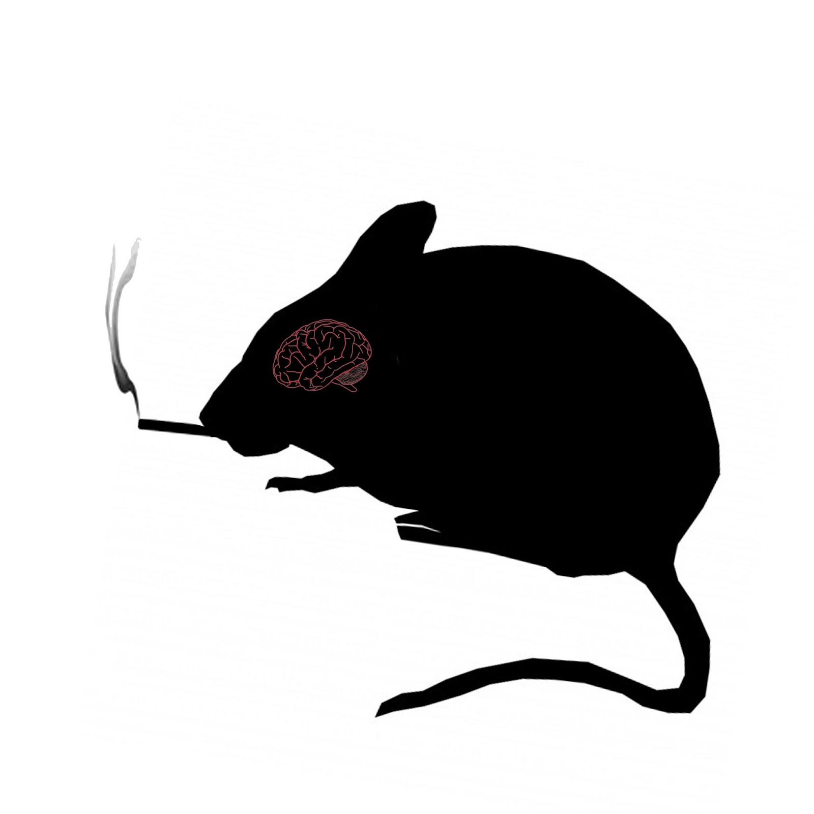
Everyone knows that nicotine can be dangerously addictive, but there’s one property of the drug that separates it from other viciously rewarding substances like cocaine and heroin. When consumed in high doses nicotine becomes acutely aversive, which helps explain why overdoses are rare compared to other drugs. So what mechanisms are responsible for this unique effect that nicotine has on the brain? And if we can better understand it, can we begin to create better tools to help people get rid of their nicotine addiction?
A team from UC Berkeley and UC Irvine constructed a computational model to begin addressing these questions. Their study focused on brain cells that produce dopamine, which plays a fundamental role in the rewarding properties of nicotine. The researchers infused high and low levels of nicotine into the blood of respective mouse groups and monitored their behavior. The mice that had received the high dose of nicotine showed less of a preference to remain in the area where the dosage had been administered, showing that they had a more aversive response to the infusion.
These behavioral results were paired with a model of cell activation that tracked the activity of dopamine producing cells in a brain region called the ventral tegmental area (VTA). The model showed a high spike in dopamine in the VTA after the first large dose of nicotine, followed by a sharp decline during subsequent doses. This drop off appears to be because high doses of nicotine desensitize acetylcholine receptors on the dopaminergic cells in the VTA, which means that the brain’s normal supply of the neurotransmitter acetylcholine is less likely to activate the cells that produce rewarding dopamine hits. Conversely, smaller doses of nicotine activate these acetylcholine receptors without drastically desensitizing them, which allows the brain to keep its dopamine production above baseline even after repeated consumption. The researchers also found a similar mechanism in the nucleus accumbens, which, alongside the VTA, is a primary brain region involved in drug response. Once again, high doses of nicotine desensitized the acetylcholine receptors that help dopaminergic neurons fire.
The team also looked into GABA producing cells and their effect on dopamine. GABA is the brain’s main inhibitory neurotransmitter, meaning that the release of GABA makes it less likely that other neurons will be activated. The researchers knew that gabaminergic neurons in the VTA and the laterodorsal tegmental nucleus connect to dopaminergic cells in the VTA and nucleus accumbens, and that if they were activated then less dopamine would be released. Sure enough, they found that aversive doses of nicotine strongly activated these GABA neurons, which correlated with an expected drop in dopamine levels.
The results of this study may help explain the neural mechanisms behind varenicline, the only drug currently available to combat nicotine addiction. The researchers hope that gaining this cellular understanding of nicotine’s unique aversion properties may contribute to even more effective treatments in the future.
Monkeys, Mice, and how Evolution Shaped Neuronal Development:
Article: “Temporally divergent regulatory mechanisms govern neuronal diversification and maturation in the mouse and marmoset neocortex”(08/01/22) - Yuan, W., Ma, S., Brown, J.R. et al. Temporally divergent regulatory mechanisms govern neuronal diversification and maturation in the mouse and marmoset neocortex. Nat Neurosci 25, 1049–1058 (2022). https://doi.org/10.1038/s41593-022-01123-4
The Rundown:
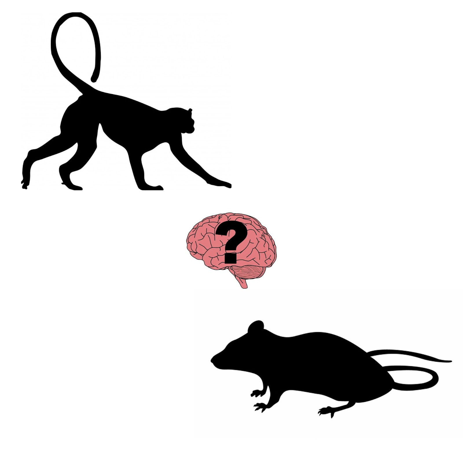
As you might already have guessed, a mouse brain is going to look pretty different from the brains of our primate cousins. You might also know that the neurons which make up these brains are some of the most diverse cell types in the body, especially in the complex brain region of the neocortex. But then again, brains are brains, and neurons are neurons, and they’re all subject to the same evolutionary laws, right? So, just how different is the developmental process of these cortical neurons across cell types and across species? And what general insights about biology can we extract from these questions?
Researchers out of Harvard University and the Max Planck Institute of Psychiatry tackled this issue by comparing different types of neocortical neurons across various stages of development in both mouse and marmoset brains. The team tracked the genetic regulation strategies of these neurons from their birth in the animal’s embryonic phase to their maturation in adulthood by measuring protein and gene expression.
They found that the early stages of neuronal development (occuring while the animals are still embryos) are more or less the same across species and neuron types when in terms of their genomic regulation. However, as they began to look at older brains in these animals, they found that the later methods of gene expression and regulation differed not only from the embryonic methods, but also that they varied far more between species and cell types. In other words, the neuronal development that occurs before birth appears to be more fundamental and consistent, holding more or less true across different species and cell types. But as the brain ages and the neurons continue to grow and form connections, we see a much greater difference in the ways that different animals and cell types express genetic regulation with regards to neuronal development.
They also found that “temporal differentiation” of regulatory strategies was common across species, meaning that both mice and marmosets use different cellular mechanisms to regulate gene expression in different stages of maturation, likely due to universal evolutionary constraints that act on all types of animals. They did, however, find differences between the species in terms of how specialized these mechanisms were to specific cell types. In maturation, mice exhibited more universal regulatory strategies that affected all neuron types indiscriminately, while the marmosets employed more differentiated strategies for the different types of neurons. This may indicate that more cognitively advanced species have a greater degree of specialization and distinction among their cortical neurons than less advanced brains.
Eating Fat Can Make You Fat, but Not for the Reasons You Think
Article:“Diet-induced inflammation in the anterior paraventricular thalamus induces compulsive sucrose-seeking” (8/01/22) - Cheng, J., Ma, X., Li, C. et al. Diet-induced inflammation in the anterior paraventricular thalamus induces compulsive sucrose-seeking. Nat Neurosci 25, 1009–1013 (2022). https://doi.org/10.1038/s41593-022-01129-y
The Rundown:
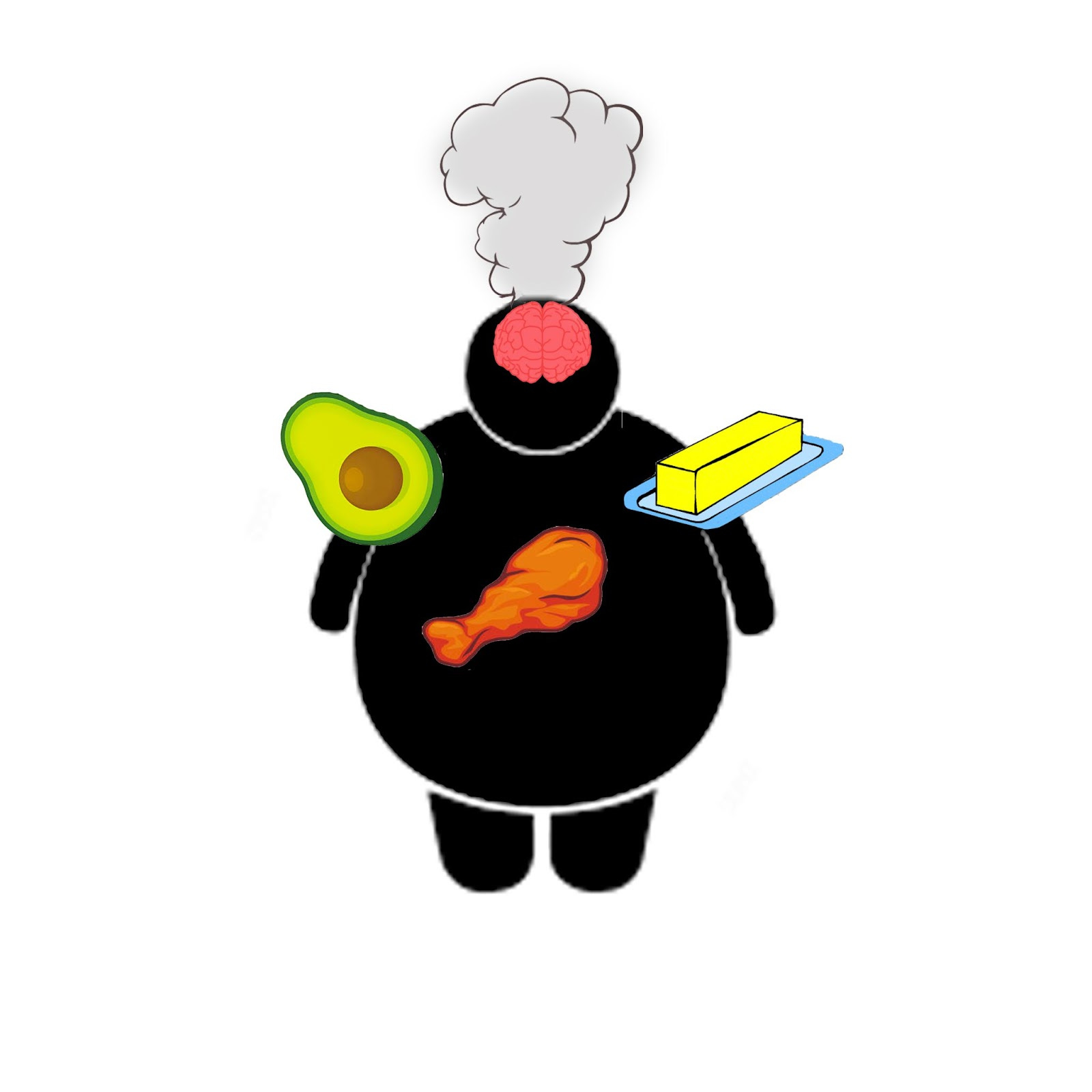
It used to be that if a person wanted to lose weight, colloquial wisdom told them to cut dietary fat first. Nowadays it seems fat is back in favor, with fatty options like the keto and Mediterranean diets gaining popularity. Recent research suggests that we may have been right the first time, at least in part. One paper demonstrates that, while fatty foods may not necessarily make you gain weight by themselves, they can impact the brain in fascinating ways that make you crave a sugary snack. Just how and why does this happen?
A team of Chinese researchers sought to dive into the intriguing effects a fatty diet has on the brain, and some of the scary implications it can have on our desire to pursue unhealthy foods in the face of risk. They used classical conditioning involving a light and an electrical shock, and measured how frequently a mouse would pursue a sugary reward in the presence of the negative conditioned stimulus. It turns out that mice which have been fed a fatty diet for a week are far more willing to withstand the risk of pain in order to receive a treat, even though they did not show a difference in pain tolerance, fear conditioning, or anxiety levels.
After measuring neuronal activation in various brain regions to find an explanation for this behavior change, the researchers found that the anterior paraventricular thalamus (aPVT) showed much more activity in high fat diet mice when it came to seeking sugar. This makes sense, since the aPVT is known to play a vital role in reward and food-seeking behavior.
Specifically, the scientists found that a mouse was more likely to seek out food under the threat of aversive stimuli when more excitatory neurons were active in the aPVT, and that this brain region seemed to be inflamed in mice that were fed a high fat diet. As an added measure of certainty, the team used optogenetics to stimulate excitatory aPVT neurons, and found that doing so made mice show similar sugar-seeking behaviors to the mice that were on a fatty diet.
A possible reason for this is that the PVT contains certain cells which monitor metabolic cues in the brain’s cerebrospinal fluid, and that inflammation in this region can disrupt the brain’s normal appetitive cues. These findings can help us better understand the neurological basis of disordered eating, and potentially help us optimize our own brains and diets to live a healthier and happier life that includes a greater control over our eating habits.
Eat more move less. I think? Ya that's the ticket
Well done!