
Neural Newsletter | Dec 2 - Dec 9
Axons, Gut-Brain Axis, and AI Stroke Detection

1. A Perspective Shift on Axonal Morphology
Publication: Membrane mechanics dictate axonal pearls-on-a-string morphology and function
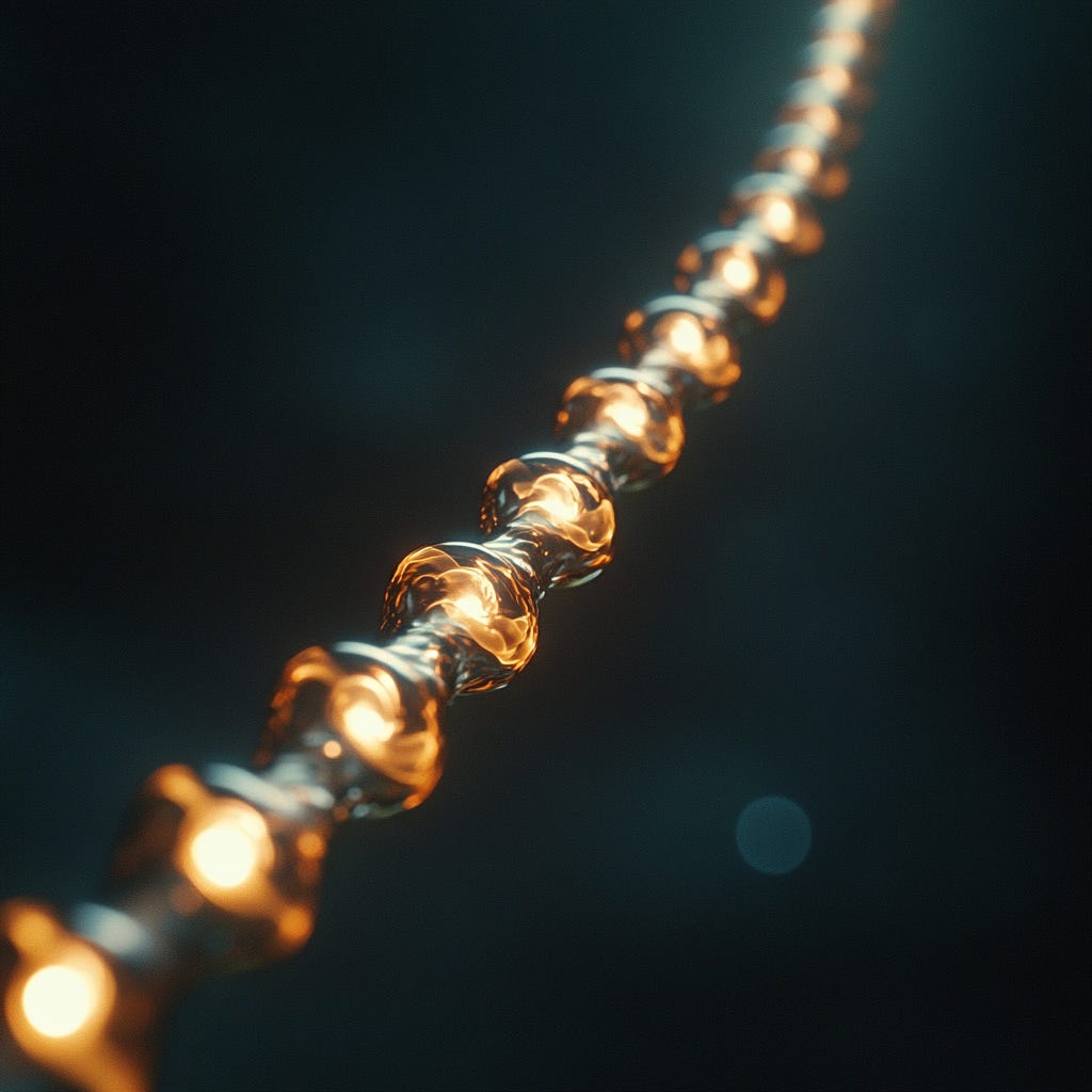
“We find that axons are not simple cylindrical tubes but rather exhibit nanoscopic pearls-on-a-string morphology due to minimization of the Helfrich–Canham energy"
- Griswold, J.M., Bonilla-Quintana, M., Pepper, R. et al., Nature Neuroscience, December 2, 2024
Anyone who has taken a high-school biology course has encountered the iconic textbook image: wispy dendrites branching from a bulbous soma, with a slender axon extending outward and splitting into root-like terminals. The axon, depicted as a simple hollow tube, serves as the link between the neuron’s integrative and transmitting ends. This basic model of axon structure has been shared not only among biology students but also among expert neuroscientists for over a century. On Monday, a study from the Watanabe lab at Johns Hopkins University came as an important reminder that no idea in science is exempt from challenge and revision, even those that appear to be right in front of our eyes.
Background, Methods, Findings
The technique at the heart of Griswold et al.’s paper is a high-pressure freezing method which, unlike classical procedures that use harsh aldehyde-based fixatives, does not distort membrane morphology. This allows for electron microscopy imaging of the axon in its near-native state. The cells used in this study included cultures of in vitro cells isolated from the hippocampus of embryonic mice, as well as acute slices from the hippocampus of adult mice.
The core finding of the paper is that neuronal axons are not simple cylinders, but instead display a beaded “pearls-on-a-string” nanostructure under normal conditions. When treated with 4% paraformaldehyde, which is common in standard fixation procedures, the cells lost this morphology within five minutes and appeared more cylindrical. The observed non-synaptic varicosities, or “nanopearls”, are ovoid bulges in the membrane about 200 nm in diameter, interspersed with thin connector regions measuring about 60 nm in diameter. This morphology is a natural product of the physical mechanics of the axonal membrane in interaction with its microenvironment. Specifically, the paper used in silico modeling and in vitro cellular manipulations to determine that osmotic conditions and membrane rigidity (modulated by cholesterol content) impact the size of nanopearls, while the cytoskeletal manipulations they performed did not.
Importantly, the morphology of these varicosities sensitively modulates the speed of action potential propagation, and seemingly functional plasticity as well. The application of 100 pulses at 100 Hz high-frequency stimulation caused an 8% increase in varicosity length and a 17% increase in width after five minutes. Electrophysiological recordings after this stimulation showed that action potential conduction was slowed, indicating a functional repercussion of this physical response.
Takeaways
This paper serves as a reminder that we should not take any of our scientific assumptions for granted. Much of our understanding of basic cellular morphology relies on observations of tissues that have been distorted by harsh fixation procedures, which can obscure the true architecture and function of cellular components in their native environment. Interestingly, C. elegans and other primitive organisms have demonstrated axonal pearling, suggesting that this is a highly conserved feature of neuronal morphology. Given that different neuronal subtypes have distinct membrane properties, which are shown here to influence the size of axonal varicosities and, consequently, the speed at which action potentials propagate along the axon, differences in membrane properties may be key determinants of functional diversity among neurons. Ultimately, Griswold et al. present a foundational and long-overlooked element of axonal morphology which reframes our understanding of neural structure and function.
2. A Functional Window into the Gut-Brain Axis
Publication: Parallel gut-to-brain pathways orchestrate feeding behaviors
“Our findings unveil parallel sensory pathways within the brainstem that encode various interoceptive signals to precisely coordinate distinct aspects of feeding regulation”
- Wang, H., Lou, R., Wang, Y. et al., Nature Neuroscience, December 3, 2024
It rarely crosses our mind while having breakfast, but this routine event poses a sophisticated challenge for the nervous system to tackle. Higher-level cues about satiation and food preferences must be processed alongside the mechanical aspects of eating, such as gulp-size, requiring the brain to integrate information from various organs. Studying how our nervous system delegates and executes this sort of foundational behavior provides a microcosmic window into the elegance of evolutionary problem-solving. The solution, described by a team at the Chinese Institute for Brain Research in Beijing, underlines the importance of specialization, integration, and parallel processing in even the most basic actions.
Background, Methods, Findings
Wang et al.’s paper is focused on the caudal nucleus of the solitary tract (cNTS), a key information hub in the lower portion of the brainstem. This region is the main integration point of information transmitted from the organs through the vagus nerve and spinal pathways, and is an important modulator of various basic behaviors including feeding. Previous work has linked several genetically distinct cell populations in the cNTS to eating behavior, but the specific function of each of individual cell groups and how they interact to coordinate feeding remains unknown. Wang et al. began by mapping the spatial distribution of different subtypes of excitatory neurons in the cNTS, which they completed using genetic tools based on published RNA sequencing data. They later performed behavioral tests, which revealed that the majority of the 19 genetically distinct neuronal populations they identified suppressed eating behavior in awake mice.
Curious about this redundancy, Wang et al. employed chemo-tagged fiber photometry to map the natural activation dynamics of nine cNTS populations during feeding. This technique involves the surgical implantation of fiber optics into the brain to measure calcium levels in specific neuronal populations, providing a readout of neural activity. This technique identified four neuronal subtypes that were highly activated by normal feeding: Gcg, Lepr, Th, and Penk. Interestingly, Gcg+ and Lepr+ neurons showed a steady increase in activation throughout the session, while Th+ and Penk+ neurons fluctuated between ~30 second pulses followed by ~1 minute gaps in activity. Because Gcg and Th are expressed by distinct cNTS neuron populations while Penk and Lepr are expressed more generally, the team chose to focus on the former to probe the functional explanations for each activation pattern.
The team found that Th+ cNTS neurons were highly activated by liquid infusions into the oral cavity, but not by infusions into the stomach. This was strong evidence that the Th population was activated by pregastric signals, but more investigation was required to determine which precise signals were linked to this population. The team found that ablating the taste buds before feeding did not change the activity of these neurons, nor did touching the oral cavity. However, prodding the esophagus induced a powerful response in Th+ neurons which could be blocked by silencing sensory transmission around the area. This indicates that this neural population is specialized for monitoring the size of gulps during feeding based on esophageal distension.
Unlike the pulsing activity patterns of Th+ neurons, the activity of Gcg+ neurons steadily increases during feeding and correlates positively with the total amount of food ingested. The team found that probing the esophagus,treating the tongue with sweet solutions, or inflating an intragastric balloon to distend the stomach did not activate the Gcg population. Instead, activity in this population correlated with the total caloric content of ingested solutions, but not the total volume of those solutions. Furthermore, they responded to the ingestion of caloric sweeteners such as sucrose, but not non-caloric sweeteners such as sucralose. These findings indicate that Gcg neurons track total caloric intake during feeding. They also found that Gcg neuron activity is modulated by the animal’s hunger, while Th neurons respond consistently regardless of internal states.
Curious about the gut-brain circuits underlying these separate behaviors, the team used viral retrograde tracing to determine where Th+ and Gcg+ cNTS neurons received inputs from. This revealed that Th+ neurons receive innervation from the vagus nerve endings lining the esophagus, while Gcg+ neurons are strongly innervated by projection neurons within the spinal afferent pathway.
Takeaways
This paper helps elucidate how diverse signals from the gut are processed in the brainstem to inform feeding behavior, which provides broader insight into how the brain encodes and synthesizes information to achieve functional outcomes. Wang et al. found that different neuronal populations are specialized to fire in response to highly specific sensory cues in the gastrointestinal tract, which reach the brain via separate pathways and converge in the cNTS to inform a cohesive behavioral response. Understanding how the brain employs specialization, parallelization, and integration reveal the principles that make it such a powerful problem-solving machine.
3. An AI-Powered Leap In Stroke Diagnosis

Publication: Deep learning biomarker of chronometric and biological ischemic stroke lesion age from unenhanced CT
“Our study shows that a CNN-radiomics based model of ischemic lesion age significantly outperforms the NWU approach. This deep learning biomarker provides more accurate estimates of stroke onset time and lesion reversibility, both critical for decisions regarding revascularization treatments.”
- Marcus, A., Mair, G., Chen, L. et al., npj Digital Medicine, December 6, 2024
Recent advances in artificial intelligence are already reshaping biomedical fields. Google DeepMind’s AlphaFold, a deep learning model for predicting protein structure, recently garnered two of its developers the Nobel Prize in Chemistry. A controversial Stanford study found that ChatGPT outperformed seasoned physicians in making diagnostic assessments from case studies, even when those physicians had access to ChatGPT. We may be on the brink of an AI revolution in medicine, and a recent study from Imperial College London indicates that neurological diagnoses could benefit. Can artificial neural networks save the biological brains of ischemic stroke patients?
Background, Methods, Findings
Estimating the age of an ischemic lesion is a critical step in stroke treatment, as it provides key insights into the lesion’s potential reversibility. The current standard approach involves measuring Net Water Uptake (NWU) from CT scans, which generates a relative intensity (RI) value. This statistic serves as an indicator of the extent of blood-brain barrier breakdown. Unfortunately, this method is imprecise and is easily confounded by biological factors and subjective misidentification of lesion sites by human experts. Marcus et al. wanted to determine whether a machine learning approach, which can identify and predict the age of ischemic lesions using variables that are imperceptible to diagnosticians, could outperform the current state-of-the-art. The resulting model combined convolutional neural networks with radiomic features to produce diagnostically-relevant biomarkers.
The team approached this problem in two stages: first, developing a method to accurately segment lesion sites from CT images, and second, using these segmented images to develop a model capable of reliably estimating lesion age. For this first task, they trained one model to identify early, acute lesions (within 6 hours), and one to identify older, subacute lesions (6-48 hours). When CT images were fed through these models, the AI system successfully identified ischemic lesions in 90.3% of cases (1,757 out of 1,945). The model’s lesion segmentation was generally in line with those of human experts, with the ASPECTS similarity score showing no significant difference between inter-expert segmentation and model-expert segmentation.
While segmentation was similar between the deep learning models and the expert diagnosticians, there was a major difference in performance when it came to estimating the age of the lesions. The R squared age estimation of the deep learning approach was 0.577 compared to 0.317 for the standard RI model, reflecting an 85% improvement. It outperformed similarly when estimating other useful features of ischemic lesions, such as core:penumbra ratio and lesion growth rates.
Takeaways
Stroke onset times are unknown in approximately 20% of cases, posing a significant challenge for determining patient eligibility for time-sensitive interventions. Accurate estimation of lesion age is critical to distinguishing salvageable tissue from irreversible damage and guiding treatment decisions. The development of a deep learning model that is nearly twice as accurate as the best existing method represents a substantial breakthrough in ischemic stroke care. By leveraging advanced imaging features, this model demonstrates that a machine learning approach can enhance diagnostic precision and equip clinicians with a robust tool to tailor interventions to individual patients. This innovation has the potential to improve outcomes for patients who lack clear symptom onset information, and also charts a path for the continued integration of artificial intelligence into medical care.
Thank you for reading this week’s Neural Newsletter! Make sure to subscribe to stay updated on recent findings in neuroscience, and click the button below to share this post with anyone who might be interested.
That axon study is pretty wild